2026-07-13 東京科学大学
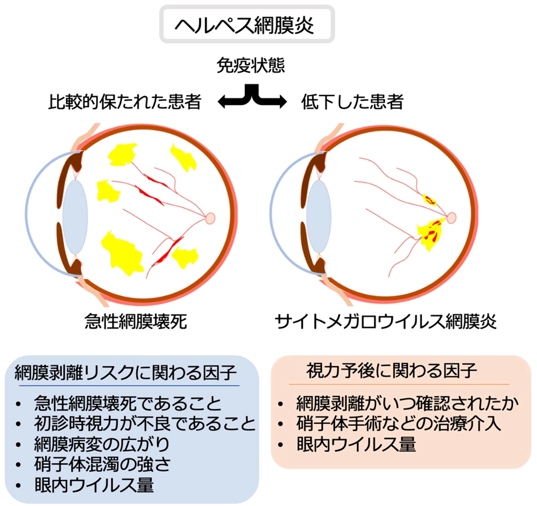
図1. ヘルペスウイルス網膜炎における重症化リスク評価
<関連情報>
- https://www.isct.ac.jp/ja/news/pnkym8uwsqeq
- https://www.sciencedirect.com/science/article/pii/S2468653026002721
免疫状態別のヘルペスウイルス網膜炎:網膜剥離および重度視覚障害の臨床表現型と予測因子
Herpesvirus Retinitis by Immune Status: Clinical Phenotypes and Predictors of Retinal Detachment and Severe Visual Impairment
Yaru Zou MD, Mingming Yang MD, Jing Zhang MD, Kyoko Ohno-Matsui MD, PhD, Koju Kamoi MD, PhD
Ophthalmology Retina Available online: 6 June 2026
DOI:https://doi.org/10.1016/j.oret.2026.06.002
Purpose
To evaluate the clinical manifestations, retinal detachment (RD) patterns, treatment strategies, and visual outcomes in herpesvirus retinitis and to identify predictors of RD and severe visual impairment.
Design
Retrospective longitudinal cohort study.
Participants
Among 1175 patients screened for suspected viral retinitis (2013–2025), 120 patients (144 eyes) with acute retinal necrosis (ARN) or cytomegalovirus retinitis (CMVR) were included.
Methods
Demographic, clinical, imaging, and treatment data were reviewed. Diagnosis was based on the Standardization of Uveitis Nomenclature classification criteria, irrespective of polymerase chain reaction (PCR) status. Multivariable regression, generalized estimating equation modeling, Kaplan–Meier survival analysis, and Cox regression were performed to compare ARN and CMVR phenotypes and identify predictors of RD and severe visual impairment (<20/200). Polymerase chain reaction testing was recorded, and subgroup analyses were performed according to PCR status.
Main Outcome Measures
Ocular and systemic characteristics between CMVR and ARN and risk factors for RD and severe visual impairment.
Results
Mean follow-up was 5.5 years. Cytomegalovirus retinitis occurred mainly in patients with lymphoma (P = 0.022) and prior immunosuppressive therapy (P = 0.027), whereas ARN more commonly showed unilateral involvement (P = 0.012), peripheral necrosis (B = 1.91; P = 0.046), and a more fulminant course (B = –0.06; P < 0.001). Retinal detachment developed earlier and more frequently in ARN (log-rank P < 0.001). In multivariable Cox analysis, ARN (hazard ratio [HR], 11.52; 95% confidence interval [CI], 1.93–68.86; P = 0.007) and worse baseline visual acuity (HR, 2.06; 95% CI, 1.23–3.44; P = 0.006) were independently associated with an increased risk of RD. Surgical intervention (B = –2.50; 95% CI, –4.72 to –0.20; P = 0.033) and concurrent RD (B = –1.28; 95% CI, –2.55 to –0.02; P = 0.047) were independently associated with lower odds of severe visual impairment. Polymerase chain reaction-based subgroup analyses showed broadly similar clinical and outcome patterns.
Conclusions
Host immune status was associated with distinct clinical phenotypes and outcome patterns in herpesvirus retinitis. Cytomegalovirus retinitis occurred in immunocompromised patients and showed a more indolent course. ARN showed broader retinal involvement and earlier RD. Retinal detachment timing and surgical intervention were associated with visual outcomes. These findings highlight distinct disease patterns and may improve early recognition and management.
Financial Disclosure(s)
Proprietary or commercial disclosure may be found in the Footnotes and Disclosures at the end of this article.