2026-07-31 カロリンスカ研究所(KI)
<関連情報>
- https://news.ki.se/somatic-mutations-linked-to-vascular-damage-in-progeria
- https://link.springer.com/article/10.1186/s13073-026-01719-6
プロジェリア動脈壁の単一細胞解析により、プロジェリン誘発性の進行性、細胞型特異的な機能障害および体細胞突然変異の蓄積が明らかになった Single-cell analysis of the progeria arterial wall reveals progerin-induced progressive, cell type-specific dysfunction and somatic mutation accumulation
Lara G. Merino,Gwladys Revêchon,Santhilal Subhash,Fabiana Stefani,Daniel Whisenant,Marianna Skipitari,Quentin Giraud,Lars Muhl,Giuseppe Mocci,Johan Björkegren,Piotr Machtel,Liqun He,Christer Betsholtz & Maria Eriksson
Genome Medicine Published:31 July 2026
DOI:https://doi.org/10.1186/s13073-026-01719-6
Abstract
Background
The premature aging disorder Hutchinson-Gilford Progeria Syndrome (HGPS) is caused by de novo LMNA mutations producing the aberrant Lamin A isoform progerin. HGPS patients die from cardiovascular disease, with their arteries showing extensive cellular and structural remodeling, but the mechanisms driving vascular dysfunction are not fully understood.
Methods
To define molecular processes underlying progressive vascular degeneration in HGPS, we performed single-cell RNA-sequencing (scRNA-seq) of aortic arch cells from LmnaG609G/G609G mice without atheroprone stimuli. These mice carry the murine equivalent of the most common HGPS-causing mutation and faithfully recapitulate the vascular phenotype. Sequencing was performed at multiple ages to capture disease-related and time-dependent transcriptional changes. We used Smart-seq2 for sequencing, due to its high sensitivity and full-length transcript coverage. Histology, immunostaining and in situ hybridization were used for arterial characterization.
Results
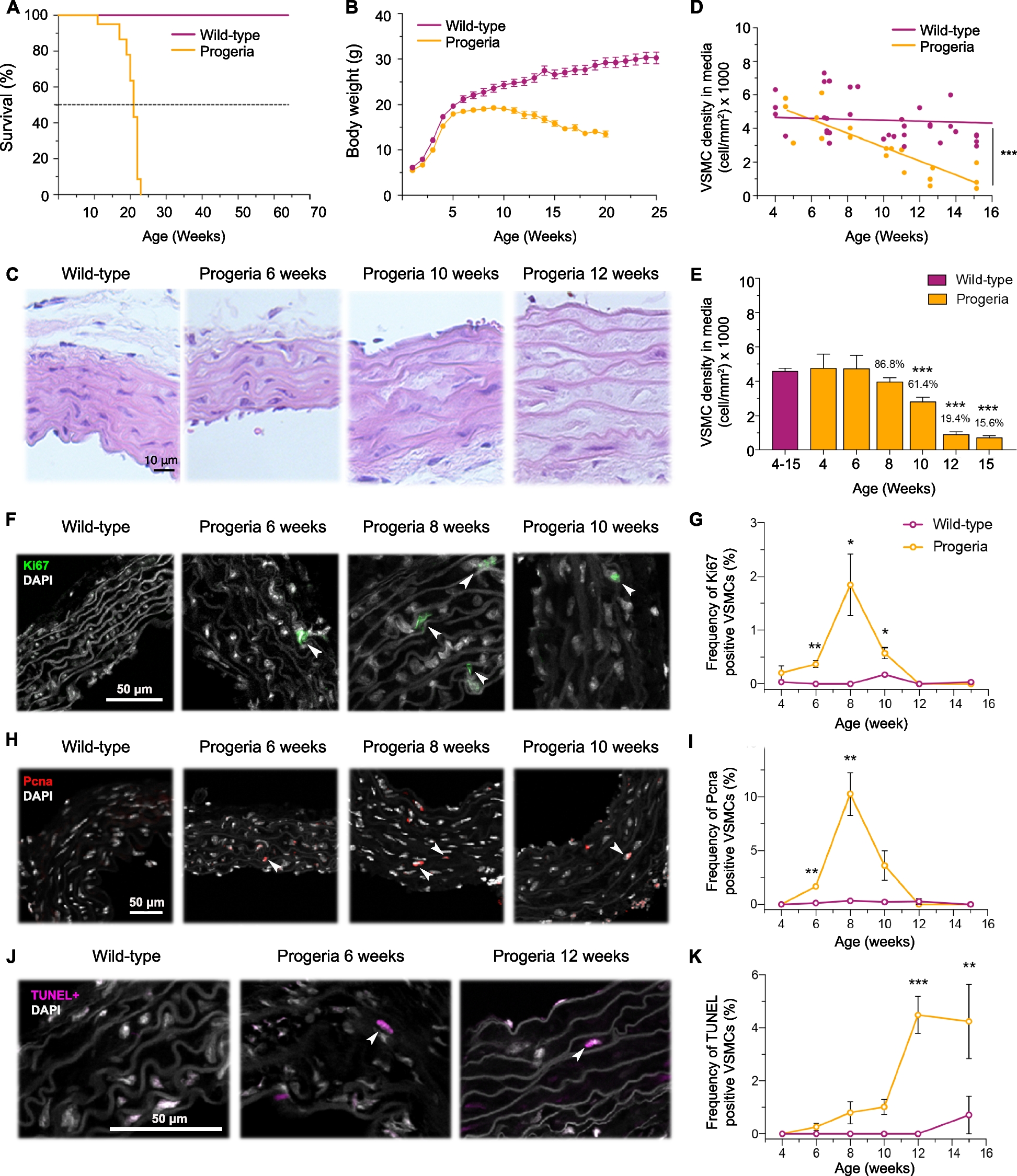
The aortic arch of LmnaG609G/G609G mice exhibited a gradual age-dependent vascular smooth muscle cell (VSMC) loss, accompanied by a transient proliferation surge, and ultimately by increased apoptosis. scRNA-seq identified transcriptionally distinct cell populations with unique features that evolved during disease progression. Disease-enriched VSMCs at early stages were characterized by elevated endoplasmic reticulum (ER) stress. With disease development, these VSMCs further underwent phenotypic switching toward a fibroblast-like state, which was predicted to expand through non-cell-autonomous mechanisms. At later stages, disease-enriched VSMCs upregulated apoptotic gene expression, partially coinciding with sustained ER stress. Furthermore, progeria VSMCs showed an increase in both DNA damage and somatic SNVs, with the increased number of SNVs correlating with high expression of ER stress, ROS and p53-related genes. In contrast, progeria-enriched fibroblasts either became activated or increased their cartilage production and showed a delayed accumulation of somatic SNVs compared to VSMCs, highlighting both a cell-type-specific progerin response and differences in somatic mutation susceptibility.
Conclusions
Our study shows that progerin leads to somatic mutation accumulation particularly in VSMCs, highlighting the need for early, cell-type-specific therapeutic intervention in HGPS to prevent permanent vascular tissue damage. In addition, the cell-type-specific molecular dynamics of the aortic arch VSMCs and fibroblasts during HGPS disease progression are provided in a user-friendly searchable scRNA-seq database available for preclinical research targeting vascular aging.