2026-10-23 東京科学大学
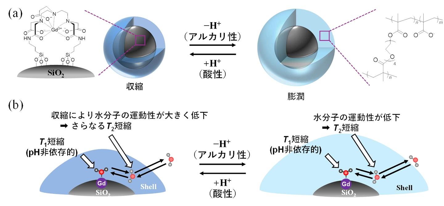
図1. 開発したコア-シェルナノ粒子の(a)模式図と、(b)予想されるT1とT2の短縮機構。
<関連情報>
生理的pHの定量的イメージングのための調整された架橋ネットワークを備えた比率測定型MRIプローブ A Ratiometric MRI Probe With a Tuned Crosslinked Network for Quantitative Imaging of Physiological pH
Ken Kato, Akira Sumiyoshi, Kyoko Katsumata, Hiroyuki Nakamura, Koichi Kato, Kazuya Kikuchi, Satoshi Okada
Small Published: 23 September 2026
DOI:https://doi.org/10.1002/smll.75796
ABSTRACT
Quantitative pH imaging is important for understanding pathological tissue environments and diagnosing diseases such as tumors and inflammation. MRI-based pH quantification has been developed based on several approaches including chemical exchange saturation transfer and ratiometric probes. However, existing approaches involve trade-offs in sensitivity, probe complexity, and reversibility, and robust quantitative pH imaging remains challenging. Previously, we reported ratiometric MRI probes based on a simple core–shell architecture in which a gadolinium-loaded core and a pH-responsive hydrogel shell independently contribute to longitudinal relaxivity (r1) and transverse relaxivity (r2), respectively. These probes enabled pH quantification using r2/r1 ratiometry although their pH responses were not optimized for the physiological pH range and their usable probe concentrations were limited. Here, we optimized the crosslinked network of the shell using tetraethylene glycol dimethacrylate, achieving substantial pH-dependent changes in r2 around physiological pH while preserving r1 and extending the usable probe concentration range. Electron microscopy confirmed the core–shell structure and elemental distribution of the probe. The resulting probe enabled quantitative pH imaging over pH 6.0–7.5 across probe concentrations from 0.5 to 1.5 wt.%. These findings establish a practical strategy for MRI-based pH quantification, offering a step toward standardized non-invasive pH measurements.