2023-10-13 ペンシルベニア州立大学(PennState)
◆これらのプロジェクトは臨床で観察される疾患に基づいており、その結果得られる発見は、将来的には皮膚疾患、がん、神経疾患などの患者に新しい治療法をもたらす可能性があります。特に、皮膚疾患を研究の文脈として使用し、細胞同士の接着や結合に関する基本的な生物学について探求しています。
◆研究者たちは、生体内で細胞同士がどのように接続し、組織を形成し、ストレスに対応するかを理解することを目指しています。その成果は、将来的に医学の進歩につながる可能性があります。
<関連情報>
- https://www.psu.edu/news/research/story/advanced-imaging-gives-researchers-front-row-view-cellular-junctions/
- https://www.nature.com/articles/s41556-023-01154-4
デスモソーム小胞体複合体の構造と動態を解明 Architecture and dynamics of a desmosome–endoplasmic reticulum complex
Navaneetha Krishnan Bharathan,William Giang,Coryn L. Hoffman,Jesse S. Aaron,Satya Khuon,Teng-Leong Chew,Stephan Preibisch,Eric T. Trautman,Larissa Heinrich,John Bogovic,Davis Bennett,David Ackerman,Woohyun Park,Alyson Petruncio,Aubrey V. Weigel,Stephan Saalfeld,COSEM Project Team,A. Wayne Vogl,Sara N. Stahley & Andrew P. Kowalczyk
Nature Cell Biology Published:08 June 2023
DOI:https://doi.org/10.1038/s41556-023-01154-4

Abstract
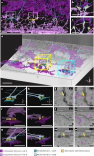
The endoplasmic reticulum (ER) forms a dynamic network that contacts other cellular membranes to regulate stress responses, calcium signalling and lipid transfer. Here, using high-resolution volume electron microscopy, we find that the ER forms a previously unknown association with keratin intermediate filaments and desmosomal cell–cell junctions. Peripheral ER assembles into mirror image-like arrangements at desmosomes and exhibits nanometre proximity to keratin filaments and the desmosome cytoplasmic plaque. ER tubules exhibit stable associations with desmosomes, and perturbation of desmosomes or keratin filaments alters ER organization, mobility and expression of ER stress transcripts. These findings indicate that desmosomes and the keratin cytoskeleton regulate the distribution, function and dynamics of the ER network. Overall, this study reveals a previously unknown subcellular architecture defined by the structural integration of ER tubules with an epithelial intercellular junction.


