2026-08-05 東北大学
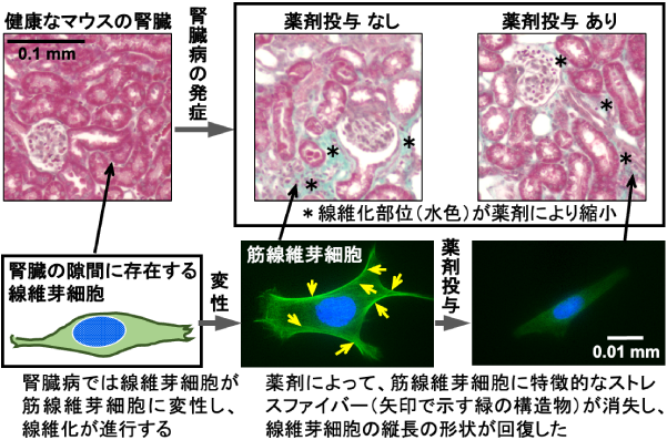
図1. 腎臓病によって線維芽細胞が筋線維芽細胞に変性し、線維化が進行する。HDAC阻害剤によって筋線維芽細胞が線維芽細胞の性質を回復し、線維化が収束するとともに、腎臓全体の障害も軽減されることが明らかとなった。
<関連情報>
- https://www.tohoku.ac.jp/japanese/2026/08/press20260805-01-myofibroblast.html
- https://www.sciencedirect.com/science/article/pii/S002432052600408X
ヒストン脱アセチル化酵素の阻害は、筋線維芽細胞の線維芽細胞様特性を回復させることで腎障害を軽減する Histone deacetylase inhibition ameliorates kidney injury by restoring fibroblast features to myofibroblasts
Hirotaka Ishioka, Yuma Iwamura, Yusuke Konta, Koji Sato, Koichiro Kato, Tetsuhiro Tanaka c, Taku Nakai, Norio Suzuki
Life Sciences Available online: 20 July 2026
DOI:https://doi.org/10.1016/j.lfs.2026.124599
Abstract
Aims
There are no effective strategies for treating chronic kidney disease (CKD) because of the limited understanding of its complex pathology. Fibrosis commonly develops and expands during CKD progression, making it a plausible therapeutic target against CKD. We have previously reported that kidney disease induces the myofibroblastic transformation of renal interstitial fibroblasts, which produce the erythroid growth factor erythropoietin, thereby promoting fibrosis through the production of extracellular matrix and leading to erythropoietin-deficiency anaemia. Since histone deacetylases (HDACs) govern cellular differentiation status, this study investigated whether HDAC inhibition could ameliorate renal fibrosis through restoring myofibroblasts to fibroblastic cells.
Methods
Changes in the gene expression profile and morphology of a myofibroblast line derived from murine renal fibroblasts were assessed after incubation with HDAC inhibitors or inactivation of HDAC isoform expression. The effects of pharmacological HDAC inhibition were also evaluated using CKD model mice and primary-cultured renal myofibroblasts obtained from the urine of a patient with CKD.
Key findings
HDAC inhibitors shifted the gene expression signature of myofibroblasts towards that of fibroblasts and induced morphological changes. HDAC2 knockdown partially recapitulated these effects, whereas HDAC1 knockdown failed because of cell death. HDAC inhibition significantly attenuated genes related to fibrosis and tubular damage, with similar trends histologically observed. In patient-derived renal myofibroblasts, HDAC inhibition suppressed profibrotic gene expression and restored erythropoietin gene expression.
Significance
HDAC inhibition suppresses renal fibrosis through the induction of apoptosis and restores fibroblast features in myofibroblasts, potentially leading to the amelioration of tubular damage and erythropoietin insufficiency in patients with CKD.