2026-08-07 近畿大学
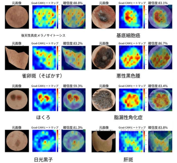
AIが「どこを見て診断したか」を示すヒートマップ
<関連情報>
- https://www.atpress.ne.jp/news/8598222
- https://www.jaadinternational.org/article/S2666-3287(26)00129-X/fulltext
説明可能なディープラーニングモデルによる臨床写真を用いた8カテゴリーの色素性皮膚病変の分類:日本における多施設共同後方視的内部妥当性検証研究
An explainable deep learning model classifies eight categories of pigmented skin lesions on clinical photographs: A multicenter retrospective internal validation study in Japan
Kimi Iinuma, MD ∙ Kazuyasu Fujii, MD, PhD ∙ Chisa Nakashima, MD, PhD ∙ … ∙ Yuichi Kimura, PhD ∙ Takashi Nagaoka, PhD ∙ Atsushi Otsuka, MD, PhD
JAAD International Published:July 23, 2026
DOI:https://doi.org/10.1016/j.jdin.2026.07.002
Pigmented skin lesions are commonly encountered in dermatologic practice, yet their differentiation based on clinical photographs remains challenging, particularly when dermoscopy is unavailable.1 We developed an explainable deep learning–based classification model for common pigmented lesions using routine clinical photographs, building upon recent advances in artificial intelligence–based skin lesion classification.2
In this multicenter retrospective study, 979 clinical photographs across 8 diagnostic categories (acquired dermal melanocytosis, basal cell carcinoma, ephelis, malignant melanoma, melasma, nevus, seborrheic keratosis, and solar lentigo) were collected from dermatologic institutions in Japan. Benign lesions were diagnosed in routine clinical practice by board-certified dermatologists using established criteria, whereas malignant melanoma and basal cell carcinoma were histopathologically confirmed. The model incorporated architectures pretrained on publicly available dermatologic image data sets.3 Data were split at the patient level into training, validation, and independent test sets to prevent data leakage, with each patient assigned to a single subset.
Class-specific receiver operating characteristic and precision–recall curves are shown in (Supplementary Fig 1, available via Mendeley at https://doi.org/10.17632/ndg49xmgpr.1). On the independent test set, the model correctly classified 188 of 196 images, yielding an accuracy of 95.9% (95% CI, 92.1%-98.2%). Melanoma sensitivity was 100% in this limited test set (20/20; 95% CI, 83.2%-100.0%), but this was not powered to establish melanoma safety. Most misclassifications occurred between clinically similar lesions, particularly seborrheic keratosis and basal