2026-07-22 東北大学
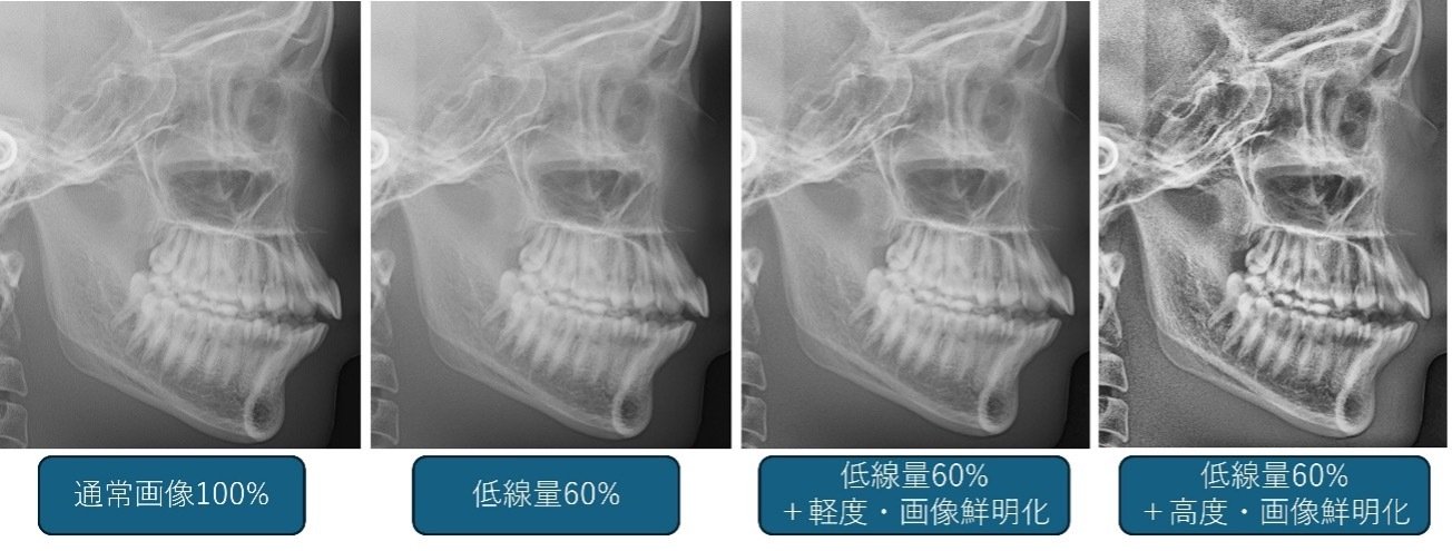
図1. 低線量側面頭部エックス線規格写真に対する画像鮮明化処理を適用
<関連情報>
- https://www.tohoku.ac.jp/japanese/2026/07/press20260722-02-cephalometric.html
- https://academic.oup.com/dmfr/advance-article/doi/10.1093/dmfr/twag047/8725343?login=false
画像鮮明化により、低線量側面頭部X線写真における基準点の視認性が向上する:ファントム研究 Image sharpening improves the visibility of reference points on low-dose lateral cephalometric radiographs: a phantom study
Ikuho Kojima, DDS, PhD,Arata Ito, DDS, PhD,Yusuke Shimada, DDS, PhD,Naoko Tagaino Watanabe, DDS, PhD,Manami Ikawa, RT,Shu Onodera, RT, PhD,Hiroyasu Kanetaka, DDS, PhD,Masahiro Iikubo, DDS, PhD
Dentomaxillofacial Radiology Published:06 July 2026
DOI:https://doi.org/10.1093/dmfr/twag047
Abstract
This phantom study aimed to determine whether a novel non-artificial intelligence image-sharpening approach improves the visibility of reference points on low-dose lateral cephalometric radiographs. Lateral cephalometric radiographs were obtained using an adult human bone phantom at 100% of the routine dose. Low-dose imaging was performed using 60, 40, and 20% of the routine dose. Each low-dose image underwent a two-level image-sharpening process using the Medical Image Enhancer software. Six reviewers evaluated the visibility of the five reference points in the first and second sets of sharpened and 100% radiation-dose images. Using Scheffé’s method of paired comparisons, odds ratios (OR) were reported as relative values for a standard cephalometric radiograph (100% radiation-dose) to compare the visibility of reference points among imaging conditions. For the first set of mildly sharpened 60%-dose images, the ORs for the four reference points were > 1.0, Point A (OR 1.12, p = 0.83), Nasion (OR 2.26, p = 0.11), posterior nasal spine (OR 2.35, p = 0.1), and incisal edge of the maxillary central incisor (OR 1.35, p = 0.53), indicating visibility comparable to or better than the 100%-dose images. For the second set of markedly sharpened images, rated visibility was significantly higher for all reference points at 60% and 40% doses and for several reference points at 20% dose. Image-sharpening algorithms improved the visibility of reference points on low-dose phantom radiographs, suggesting their technical feasibility as a post-acquisition approach for dose-reduced cephalometric imaging.