2026-07-22 国立環境研究所,大阪大学,早稲田大学
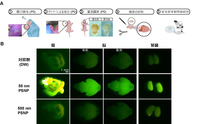
図1 新生仔マウスに経口投与した蛍光ポリスチレン製ナノプラスチックのサイズ依存的生体内分布
<関連情報>
- https://www.nies.go.jp/pr/news-and-updates/2026/Press20260722-1.html
- https://www.sciencedirect.com/science/article/pii/S2772416626003499
新生マウス脳におけるナノプラスチックの可視化のための組織全体分布解析 Whole-tissue distribution analysis for visualization of nanoplastics in the neonatal mouse brain
Yang Mi, Tomohiro Ito, Kosuke Tanaka, Osamu Udagawa, Masaki Kakeyama, Yasuo Tsutsumi, Fumihiko Maekawa
Journal of Hazardous Materials Advances Available online: 29 June 2026
DOI:https://doi.org/10.1016/j.hazadv.2026.101353
Highlights
- Oral nanoplastic exposure shows size-dependent biodistribution in neonatal mice.
- Optical clearing and LSFM enable whole-brain 3D mapping of nanoplastic uptake.
- Regional brain accumulation was highest after 50 nm PS exposure.
- Workflow preserves anatomical integrity for quantitative biodistribution analysis.
Abstract
Nanoplastics (NPs) are increasingly recognized as environmental contaminants and have been detected in diverse biological tissues. However, their biodistribution in complex organs remains poorly understood. Conventional section-based imaging restricts spatial context and volumetric analysis, making it challenging to map NP distribution in structurally complex organs such as the brain even with fluorescent labeling. To address these limitations, we developed an integrated workflow combining tissue optical clearing (SeeDB2G) with light-sheet fluorescence microscopy (LSFM). This approach enables three-dimensional visualization of fluorescently labeled polystyrene (PS) NPs in neonatal mouse brains. At postnatal day 0—a critical window of heightened vulnerability due to immature barrier systems and rapid neurodevelopment—pups were orally administered spherical fluorescent 50 nm or 500 nm PSNPs under a high-dose proof‑of‑concept model designed to ensure robust whole-organ fluorescence detection. Fluorescence stereoscopic imaging revealed pronounced organ-level accumulation of 50 nm PSNPs in the intestine, liver, kidney, and brain, compared with markedly lower signals from 500 nm PSNPs 24 h post-exposure. Optical clearing rendered the entire brain transparent with preservation of fluorescence, allowing LSFM to accurately assess regional PSNP accumulation without sectioning. This workflow enabled whole-brain visualization of size-dependent NP uptake, with 50 nm PSNP detected throughout the brain and the highest relative fluorescence signals observed in the thalamus and brainstem. Although the current implementation relies on fluorescent labeling and model NPs, this approach provides a scalable platform for whole-organ biodistribution analysis and lays the foundation for mechanistic studies of barrier permeability, developmental vulnerability, and organ-specific interactions.