2026-10-09 東京大学
ゴルジ体由来小胞による新たなミトコンドリア融合機構
<関連情報>
- https://www.rcast.u-tokyo.ac.jp/ja/news/release/20261009.html
- https://www.science.org/doi/abs/10.1126/science.adx9628
PI(3,4) P2を含むゴルジ体由来小胞がミトコンドリア融合を促進する Golgi-derived vesicles containing PI(3,4)P2 drive mitochondrial fusion
Sho Aki, Mayuko Segawa, Vincent Anton, Suvagata R. Chowdhury, […] , and Julien Prudent
Science Published:8 Oct 2026
DOI:https://doi.org/10.1126/science.adx9628
Structured Abstract
INTRODUCTION
Mitochondria are double-membrane–bound organelles that generate adenosine triphosphate (ATP) and form a dynamic network remodeled by cycles of fusion and fission. These shape transitions are essential for organelle function, metabolic adaptation, mitochondrial quality control, and cell fate, and defects in these processes are associated with human disease. Although the functions of the core guanosine triphosphatases (GTPases) that drive mitochondrial dynamics are well established, these processes are now recognized to be coordinated by other organelles and regulated by local lipid signaling. However, beyond the endoplasmic reticulum (ER), whether and how other organelles, in particular, Golgi-derived vesicles, regulate mitochondrial fusion remain understudied.
RATIONALE
Phosphoinositides are low-abundance signaling lipids that are compartmentalized and regulate membrane integrity, trafficking, and remodeling. Phosphatidylinositol 4-phosphate [PI(4)P]–containing Golgi-derived vesicles facilitate mitochondrial division, raising the possibility that distinct vesicle populations containing different phosphoinositides may also regulate mitochondrial fusion. Confocal microscopy analysis of the phosphoinositide family revealed that phosphatidylinositol 3,4-bisphosphate [PI(3,4)P2]–positive structures were closely associated with mitochondria. PI(3,4)P2 could be generated from PI(4)P by the class II phosphatidylinositol 3-kinases (PI3Ks). We hypothesized that class II PI3Ks and PI(3,4)P2-containing Golgi-derived vesicles may contribute to mitochondrial dynamics and, in particular, to mitochondrial fusion. To address this, we studied the effects of genetic alterations of PI3K-C2α and/or PI3K-C2β (PI3K-C2α/β) on mitochondrial morphology and examined the organelle dynamics that occur at mitochondrial fusion sites, combining confocal microscopy, live-cell super-resolution imaging, biochemical assays, cell-based and in vitro mitochondrial fusion assays, and in vivo analyses.
RESULTS
Loss of PI3K-C2α/β, or of their kinase activity, caused mitochondrial fragmentation in mammalian cell lines. This phenotype resulted from impaired mitochondrial fusion rather than increased division. Accordingly, PI(3,4)P2-positive Golgi-derived vesicles were recruited to bona fide mitochondrial fusion events marked by the ER and the outer mitochondrial membrane (OMM) fusion proteins mitofusin 1 and 2 (MFN 1 and 2). Additionally, PI(3,4)P2 promoted mitochondrial fusion in vitro, supporting a direct role for PI(3,4)P2 in the fusion process. Investigation of the interplay between PI(3,4)P2 and the core OMM fusion machinery revealed no direct interaction between mitofusin 2 and PI(3,4)P2 but showed that loss of PI3K-C2α/β increased MFN1 and 2 oligomer accumulation, suggesting that this regulation occurs during late stages of OMM fusion.
Monitoring mitochondrial fusion and fission events revealed that distinct Golgi-derived vesicle populations were associated with different mitochondrial remodeling events. Whereas PI(4)P-positive vesicles were present at fission sites, PI(4)P and PI(3,4)P2 double-positive vesicles were associated with fusion events, indicating that PI3K-C2α/β–dependent conversion of PI(4)P to PI(3,4)P2 generates a subset of vesicles that regulate mitochondrial fusion. Exploring the physiological relevance of this process, we found that PI3K-C2α and PI3K-C2β loss in a cardiomyocyte-specific PI3K-C2α and PI3K-C2β double-deletion mouse model led to mitochondrial fragmentation and caused severe cardiac defects and early lethality, further highlighting the importance of this regulation in vivo.
CONCLUSION
Our study identifies PI(3,4)P2-positive Golgi-derived vesicles as regulators of mitochondrial fusion and class II PI3Ks as important for cardiac function in vivo. These findings support a model in which cells use specific phosphoinositide-containing Golgi-derived vesicles to coordinate either mitochondrial fusion or fission.

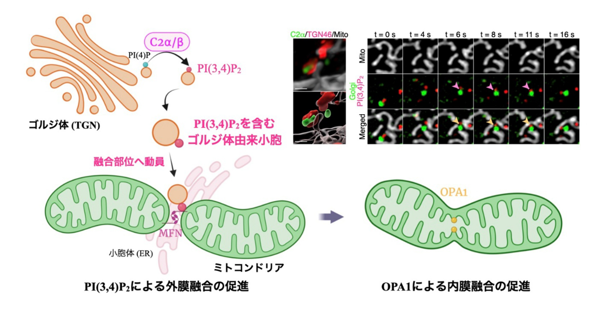
PI(3,4)P2-containing Golgi-derived vesicles coordinate mitochondrial fusion.
A subset of Golgi-derived vesicles containing PI(3,4)P2 that was generated from PI(4)P by class II PI3Ks is recruited to ER- and mitofusin-marked mitochondrial fusion sites, facilitating fusion. On the left are super-resolution images of HeLa cells showing mitochondria (visualized with TOM20), Golgi-derived vesicles (TGN46), and PI(3,4)P2 (PHTAPP1); on the right is a simplified schematic representation of the mitochondrial fusion process. Yellow arrows indicate PI(3,4)P2-positive Golgi-derived vesicles in close association with mitochondria at potential fusion sites. Scale bar is 2 μm. IMM, inner mitochondrial membrane.
Abstract
Mitochondria are dynamic organelles that remodel their shape to regulate cell fate. Mitochondrial division involves interactions with the endoplasmic reticulum (ER), lysosomes, and trans-Golgi network–derived vesicles to facilitate membrane scission. How interorganelle contacts regulate mitochondrial membrane fusion remains largely unknown. Here, we identified a role for Golgi-derived vesicles enriched in phosphatidylinositol 3,4-bisphosphate [PI(3,4)P2] in regulating mitochondrial fusion. We found that these vesicles were recruited to ER- and mitofusin-marked fusion sites. Accordingly, loss of class II PI3-kinase isoforms α and β (PI3K-C2α and PI3K-C2β), which generate PI(3,4)P2, led to mitochondrial fragmentation resulting from impaired fusion. Furthermore, cardiomyocyte-specific PI3K-C2α and PI3K-C2β double-deletion mice exhibited mitochondrial fragmentation and heart failure. Thus, subpopulations of Golgi-derived vesicles carrying different phosphoinositides control mitochondrial membrane remodeling and homeostasis.