2026-09-03 成育医療研究センター
<関連情報>
- https://www.ncchd.go.jp/press/2026/0903.html
- https://www.sciencedirect.com/science/article/pii/S1323893026000900
マウスにおいて、コハク酸の継続投与はタフト細胞-IL-25経路を介して好酸球性腸炎を誘発する Continuous succinate administration induces eosinophilic enteritis via the tuft cell-IL-25 axis in mice
Ryo Matsuoka, Yuka Hayashi, Naoko Nagano, Hiroki Sugiyama, Ayako Yamada, Keisuke Orimo, Masato Tamari, Kenichiro Motomura, Satoshi Fujita, Hisataka Nakazaki, Hiromichi Yamamoto, Taro Higuchi, Masashi Ikutani, Ken Arae, Kiyoshi Yamaguchi, Yoichi Furukawa, Katsuko Sudo, Kimihiko Oishi, Hirohisa Saito, Susumu Nakae…Kenji Matsumoto
Allergology International Available online: 22 August 2026
DOI:https://doi.org/10.1016/j.alit.2026.08.002
Abstract
Background
Eosinophilic gastrointestinal disorders (EGIDs), excluding eosinophilic esophagitis (EoE), are increasingly recognized conditions with poorly understood pathogenesis. Current animal models inadequately represent the complexity of human non-EoE EGIDs, thereby limiting therapeutic development. We aim to establish a new mouse model of eosinophilic enteritis (EoN) using continuous succinate administration and elucidate the underlying immunological mechanisms.
Methods
Four-week-old wild-type C57BL/6J mice received succinate (150 mM) in drinking water for 21 days. We analyzed eosinophil infiltration, tuft cell populations, body weight changes, and cytokine expression. Knockout mice deficient in Pou2f3 (Skn-1a), Il25, Il5, Il13, and Rag2 were used to investigate mechanistic pathways. Transcriptomic analysis compared gene expression patterns between our model and human EGID datasets.
Results
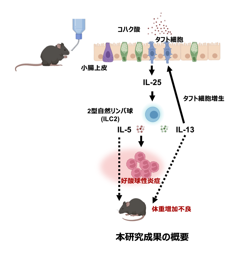
Continuous succinate administration induced significant eosinophilic infiltration in the ileum (p < 0.05), accompanied by tuft cell proliferation and failure to thrive (p < 0.001). These changes occurred without neutrophil infiltration, elevated IgE levels, or diarrhea. Pou2f3 knockout mice, lacking the transcription factor required for tuft cell differentiation, showed complete abrogation of both eosinophilic inflammation and failure to thrive, demonstrating tuft cell dependency. IL-25 and IL-5 deficiency prevented eosinophilic infiltration and failure to thrive, whereas IL-13 knockout specifically blocked tuft cell proliferation. Transcriptomic analysis revealed overlap with human eosinophilic colitis gene expression patterns, particularly in IL-13 and serotonin receptor pathways.
Conclusions
We established a new mouse model of eosinophilic enteritis recapitulating key features of human non-EoE EGIDs through the tuft cell-IL-25-ILC2 axis. This model provides new insights into EGID pathogenesis and offers a valuable platform for therapeutic development.