2026-08-27 東京大学
<関連情報>
- https://www.ims.u-tokyo.ac.jp/imsut/jp/about/press/page_00413.html
- https://www.cell.com/iscience/fulltext/S2589-0042(26)02615-5
特発性ネフローゼ症候群におけるリツキシマブ反応性に関連するT細胞の単一細胞トランスクリプトームシグネチャー Single-cell transcriptomic signatures of T cells associated with rituximab responsiveness in idiopathic nephrotic syndrome
Eri Koshi-Ito ∙ Yu Watanabe ∙ Chikao Onogi ∙ … ∙ Kazuhiro Furuhashi ∙ Shoichi Maruyama, ∙ Hiroshi I. Suzuki
iScience Published:August 21, 2026
DOI:https://doi.org/10.1016/j.isci.2026.117237
Highlights
- T cell signatures differentiate RTX responders from non-responders in MCD (INS)
- Responders show RTX-driven broad transcriptomic changes and distinct ROS regulation
- CD4+ cytotoxic T cells undergo clonal expansion with enhanced OXPHOS in responders
- Transcriptomic changes by RTX are modest in non-responders of steroid-dependent MCD
Summary
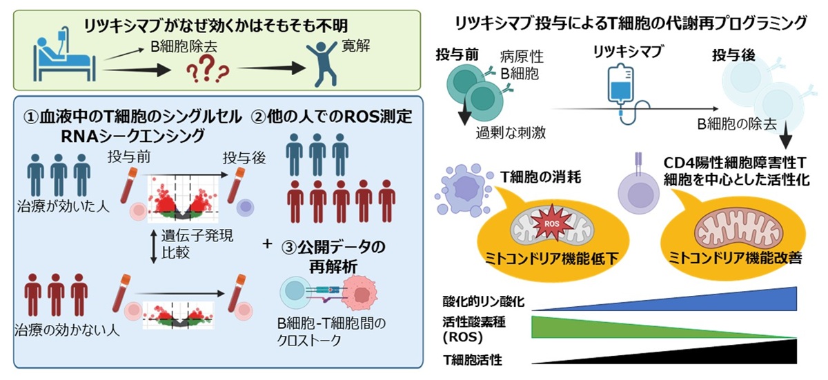
Rituximab (RTX) is increasingly used in steroid-dependent minimal change disease, a leading cause of idiopathic nephrotic syndrome (INS), yet predictors of response remain unclear. Although RTX primarily targets B cells, T cells are also implicated in INS pathogenesis. We investigated RTX-induced T cell dynamics by single-cell RNA sequencing/T cell receptor profiling of peripheral T cells from three responders and three non-responders before and after RTX treatment. Responders exhibited RTX-driven broad transcriptomic remodeling, accompanied by reduced exhausted CD8+ T cells and clonal expansion of CD4+ cytotoxic T cells with enhanced oxidative phosphorylation (OXPHOS) signatures, whereas non-responders showed marginal changes. In a separate cohort, reactive oxygen species (ROS) levels tended to be higher in responders, and declined post-RTX, collectively associating enhanced mitochondrial activity with favorable response. Reanalysis of childhood INS data supported altered B–T cell crosstalk and T cell metabolism. Together, T cells may influence RTX responsiveness, warranting further biomarker studies.