2026-11-13 金沢大学,科学技術振興機構
<関連情報>
- https://www.jst.go.jp/pr/announce/20261113/index.html
- https://www.jst.go.jp/pr/announce/20261113/pdf/20261113.pdf
- https://www.nature.com/articles/s41467-026-78964-1
泡状幾何学的不安定性によって誘導される核形成と成長を通じた上皮組織の多層化(Epithelium stratifies via nucleation and growth induced by foam-geometric instability)
Shuya Fukamachi, Razib Datta, Duligengaowa Wuergezhen, Takehiko Ichikawa, Rei Yagasaki, Phoebe Leeaw, Makiko Arai, Aki Teranishi, Shuhei A. Horiguchi, Kohei Omachi, Aiko Sada, Itaru Imayoshi, Kentaro Kajiwara, Tetsuya Hiraiwa, Masanobu Oshima, Takeshi Fukuma, Hironobu Fujiwara, Satoru Okuda
Nature Communications, Volume 17, Article 11418 (2026)2026-11-13
DOI:10.1038/s41467-026-78964-1
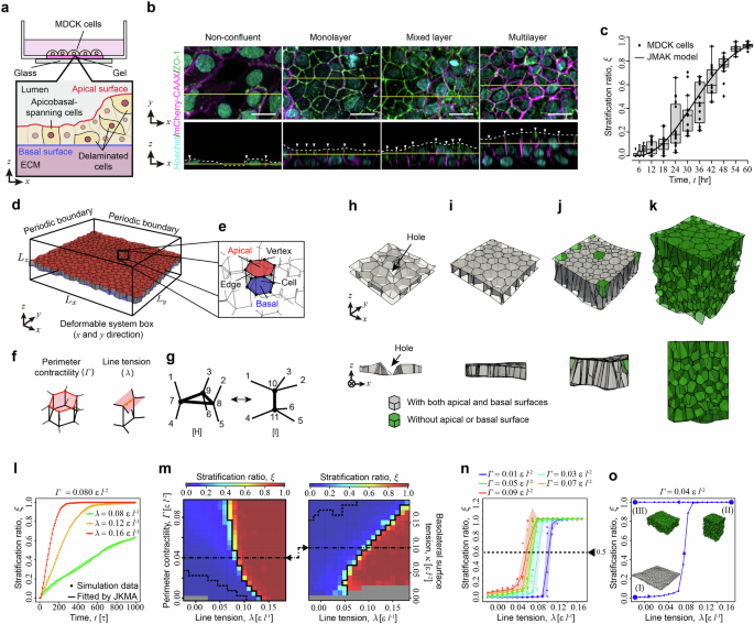
Fig. 1: Epithelial cells stratify through a nucleation-growth process. Source: Fukamachi et al., Nature Communications 17, 11418 (2026). https://doi.org/10.1038/s41467-026-78964-1. CC BY 4.0.
上皮組織が単層から多層へ変化する仕組みを明らかにするため、細胞培養実験と三次元頂点モデルを組み合わせ、上皮の多層化を解析した。MDCK細胞の培養では、単一細胞の層からの離脱が組織内の複数箇所で独立して発生する「核形成」と、発生した多層領域の境界で離脱が進む「成長」の二段階で、多層構造が広がることを示した。この過程は、泡状に詰まった上皮細胞の三次元幾何学に内在する力学的不安定性によって誘導される。細胞密度や頂端側の接合部張力の増加、隣接細胞数の減少などが細胞離脱を促し、細胞間接触の再編成にはスクートイドと呼ばれる三次元的な細胞形状が関与した。さらに、マウス胚の皮膚発生と腸管がんオルガノイドの悪性化過程でも、核形成と成長に類似した多層化が観察された。本研究は、上皮の多層化を個々の細胞の挙動だけでなく、組織全体の構造転移として捉え、発生やがん化に共通する力学的原理を提示した。なお、細胞培養・数理モデルを中心とした研究であり、組織ごとの具体的な制御機構にはさらなる検証が必要である。
To clarify how epithelial tissues transition from a monolayer to a multilayer structure, this study combined cell-culture experiments with three-dimensional vertex modeling. Experiments using MDCK cells revealed that stratification proceeds through two stages: nucleation, in which individual cells delaminate independently at multiple locations, and growth, in which delamination progresses along the boundaries of existing multilayer regions. This process is driven by mechanical instability inherent in the three-dimensional, foam-like geometry of epithelial tissues. Increased cell density, elevated apical junctional tension, and a reduction in the number of neighboring cells can promote cell delamination. Scutoids, three-dimensional cell shapes associated with rearrangement of cell–cell contacts, also contribute to this process. Similar nucleation-and-growth patterns were observed during mouse embryonic skin development and malignant transformation of intestinal cancer organoids. The findings conceptualize epithelial stratification as a tissue-level structural transition and suggest a shared mechanical principle underlying developmental and pathological epithelial remodeling. However, the study relies substantially on cell-culture experiments and mathematical modeling, and further investigation is needed to clarify the specific regulatory mechanisms in different tissues.