2026-05-28 沖縄科学技術大学院大学
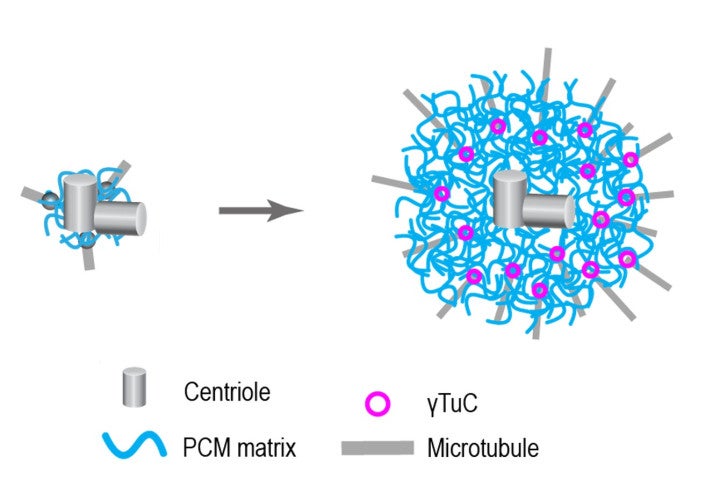
細胞分裂の際、中心体周辺物質(PCM)(青色の線)が拡張する。線虫C. elegansにおいて、中心体マトリックスの主要な構造タンパク質はSPD-5であり、SPD-5が活性化されるとγーチューブリン複合体(紫色の円)に結合する。その後、γーチューブリン複合体は微小管(灰色の線)が形成される場所として機能し、紡錘糸の形成につながる。© 太田緑(OIST)
<関連情報>
- https://www.oist.jp/ja/news-center/news/2026/5/21/scientists-reveal-how-dividing-cells-precisely-trigger-spindle-formation
- https://www.science.org/doi/10.1126/sciadv.aed6539
リン酸化は有糸分裂中心体マトリックスを再構築し、二分割γ-チューブリン複合体ドッキング部位を生成する Phosphorylation remodels the mitotic centrosome matrix to generate bipartite γ-tubulin complex docking sites
Midori Ohta, Orie Arakawa, Yajie Gu, Wanying Tian, […] , and Karen Oegema
Science Advances Published:27 May 2026
DOI:https://doi.org/10.1126/sciadv.aed6539
Abstract
Mitotic centrosomes consist of centrioles surrounded by a proteinaceous matrix that docks and activates γ-tubulin complexes (γTuCs) to nucleate microtubules for spindle assembly. During mitotic entry, phosphorylation at centrosomes remodels CDK5 regulatory subunit associated protein 2 (CDK5RAP2) family matrix proteins to generate γTuC docking sites. We address the mechanism of this conversion using Caenorhabditis elegans SPindle Defective (SPD-5) as a model. We show that SPD-5 contains two regions, phospho-regulated γTuC binding region 1 (PRGB1) and PRGB2, that are each sufficient for polo-like kinase 1 (PLK1) phosphorylation–regulated γTuC binding. We define key phosphosites in each region and uncover autoinhibition mediated by interactions within and between them. PRGB2 is dimeric and requires γTuCs containing the Mozart family microprotein MZT-1 for binding, whereas PRGB1 is monomeric and binds independently of MZT-1. Our results support a model in which PLK1 phosphorylation induces a conformational change that enables MZT-1–dependent PRGB2 engagement, which in turn relieves PRGB1 inhibition. Such a multistep mechanism would ensure robust spindle assembly by restricting microtubule nucleation in space and time.