
2026-05-22 米国国立衛生研究所(NIH)
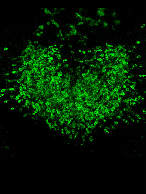
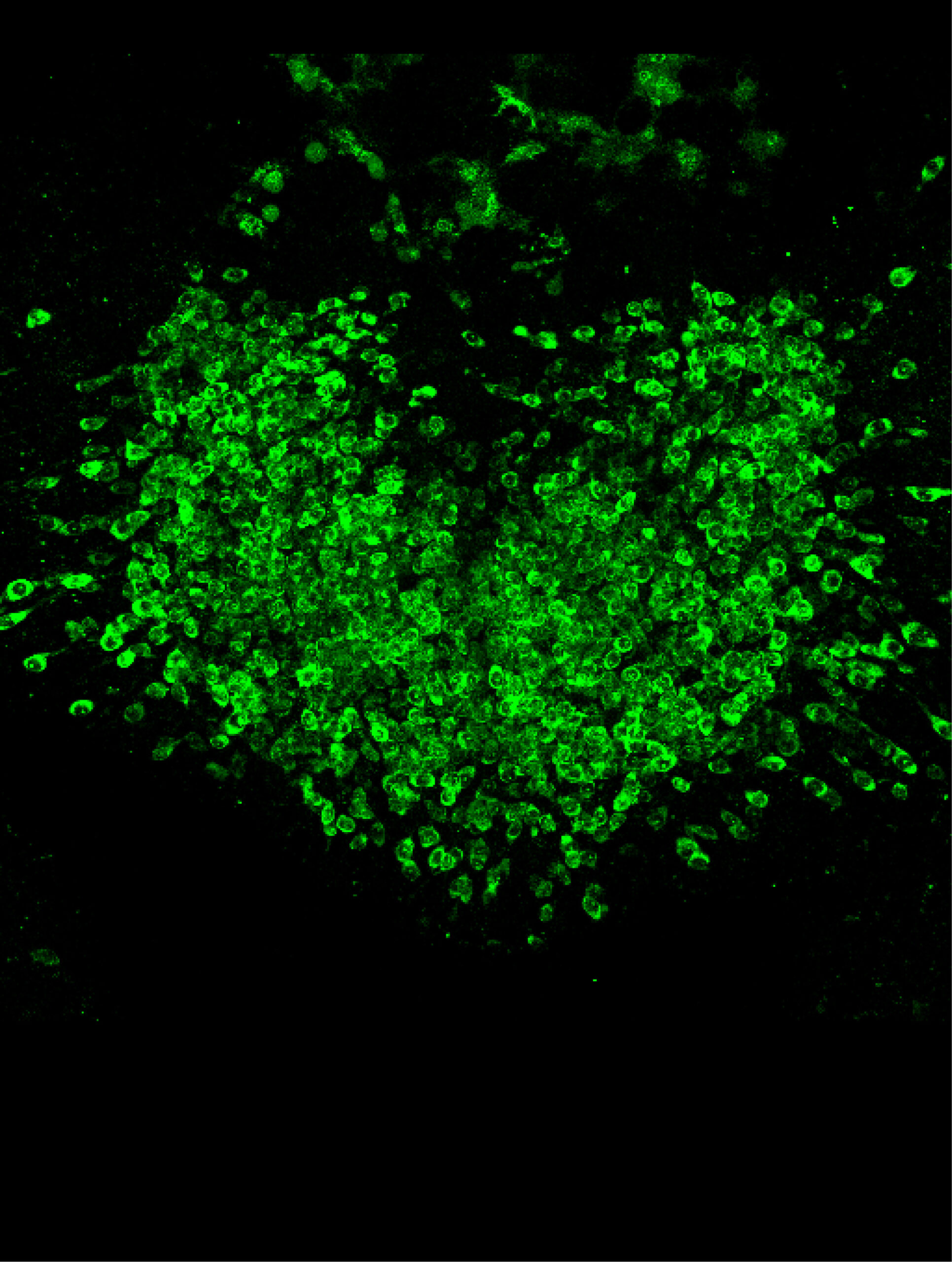
A microscopy image illustrates the varied levels of intracellular calcium responses (green) to semaglutide across GLP-1R-expressing neurons within the area postrema of the brain.
<関連情報>
- https://www.nih.gov/news-events/news-releases/nih-researchers-identify-avenue-enhanced-glp-1-induced-weight-loss
- https://www.nature.com/articles/s42255-026-01534-8
セマグルチドは、GLP1Rを発現する後脳ニューロンにおけるcAMP依存性メカニズムを介して体重減少を促進する Semaglutide drives weight loss through cAMP-dependent mechanisms in GLP1R-expressing hindbrain neurons
Claire Gao,Isabelle C. Geneve,Shakira Rodriguez-Gonzalez,Chia Li,Kaitlyn McElhern,Marc L. Reitman,Andrew Lutas & Michael J. Krashes
Nature Metabolism Published:22 May 2026
DOI:https://doi.org/10.1038/s42255-026-01534-8
Abstract
Glucagon-like peptide 1 receptor (GLP1R) agonists, such as semaglutide, drive weight loss by binding to GLP1Rs—classically described as Gs-coupled G-protein-coupled receptors—in the brain; however, the intracellular signalling mechanisms underlying these effects remain poorly defined. Here, we find that semaglutide engages both Gs– and Gq-dependent signalling pathways in Glp1r-expressing neurons in the area postrema (APGlp1r), the primary site of semaglutide action in the brain, and differentially regulates neuronal activation across distinct neuronal clusters. Semaglutide also drives graded increases of the essential secondary messenger cyclic adenosine monophosphate (cAMP) in APGlp1r neurons through the Gs pathway. Inhibition of the cAMP-degrading enzyme phosphodiesterase 4 (PDE4) enhances and sustains these cAMP responses, and disruption of Gs or cAMP signalling in APGlp1r neurons abolishes semaglutide-induced weight loss and downstream brain-wide activation. Our systematic characterization of semaglutide’s signalling mechanisms in the hindbrain reveals the intracellular signalling architecture through which semaglutide engages cAMP and calcium to regulate body weight, providing avenues for improving obesity therapeutics.

