2026-07-01 中国科学院(CAS)
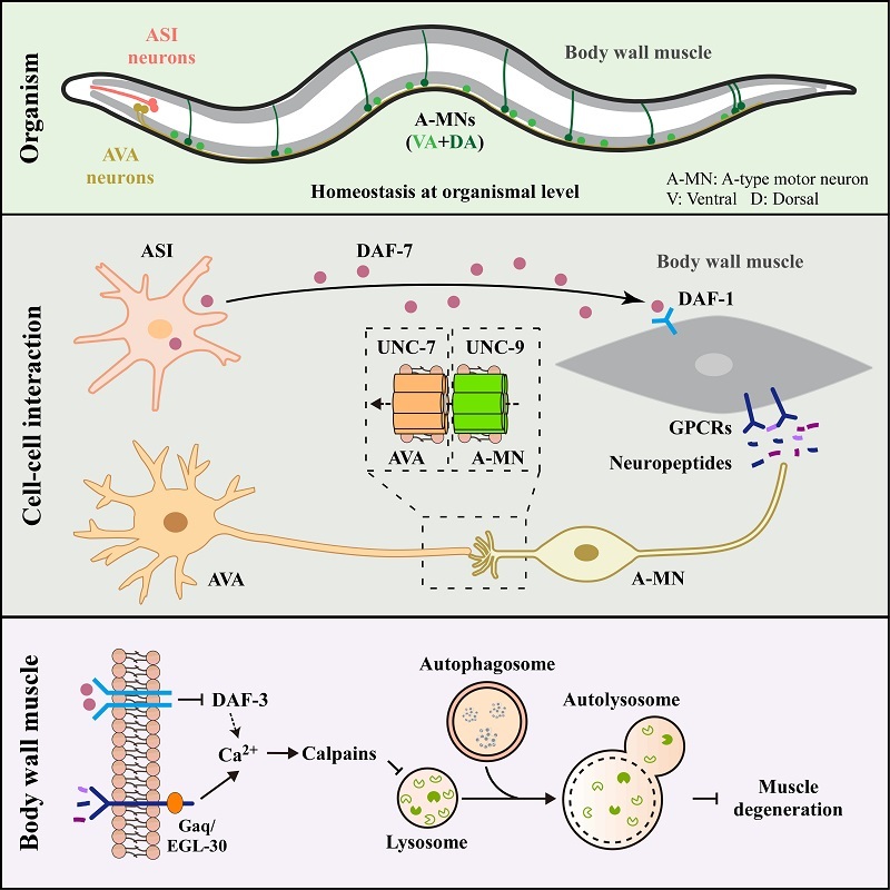
Model for how neuron-to-muscle singling orchestrates muscle autophagy and homeostasis (Image by ZHANG Hong’s group)
<関連情報>
- https://english.cas.cn/newsroom/research-news/202607/t20260702_1175221.shtml
- https://www.sciencedirect.com/science/article/abs/pii/S1534580726002182
電気シナプスとDAF-7/TGF-βシグナル伝達を含む2つの並列神経回路が、線虫C. elegansの筋肉オートファジーを制御する Two parallel neuronal circuits involving electrical synapse and DAF-7/TGF-β signaling regulate muscle autophagy in C. elegans
Hui Zheng, Hongyu Zhao, Hong Zhang
Developmental Cell Available online: 29 June 2026
DOI:https://doi.org/10.1016/j.devcel.2026.06.001
Highlights
- Two parallel neuronal circuits coordinate autophagy in C. elegans body wall muscle
- AVA/A-MN electrical synapses promote autophagy by inhibiting neuropeptide release
- ASI-secreted DAF-7/TGF-β activates autophagy via canonical TGF-β signaling
- Circuits maintain muscle Ca2+ and lysosome function to prevent neurogenic myopathy
Summary
The systemic coordination of autophagy during development remains poorly understood. Here, we identify two parallel neuronal circuits that regulate the autophagy-lysosome pathway in the body wall muscle of C. elegans. One circuit, utilizing UNC-7/UNC-9 electrical synapses between AVA interneurons and A-type motor neurons (A-MNs), promotes autophagy by inhibiting neuropeptide release from A-MNs. The other employs the TGF-β-like molecule DAF-7, secreted from ASI sensory neurons, which activates autophagy via the canonical TGF-β pathway. These pathways converge to regulate cytosolic Ca²⁺ levels in the muscle, thereby maintaining lysosomal integrity. Disruption of either circuit elevates Ca²⁺, overactivating calpain. This leads to the accumulation of non-degradative autolysosomes and accelerates muscle degeneration. Our findings elucidate a neuronal mechanism for controlling muscle autophagy and provide insights into the pathogenesis of neurogenic myopathy.