2026-08-28 京都大学
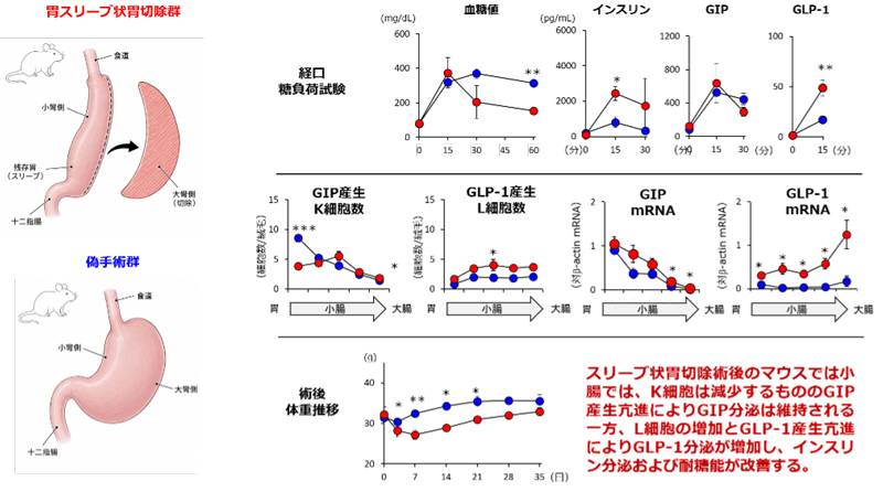
本研究成果の概要
<関連情報>
- https://www.kyoto-u.ac.jp/ja/research-news/2026-08-28
- https://journals.bioscientifica.com/jme/article-abstract/doi/10.1530/JME-26-0016/78059/An-analysis-of-intestinal-morphology-and-incretin
雄マウスにおけるスリーブ状胃切除術後の腸管形態およびインクレチン産生細胞の解析 An analysis of intestinal morphology and incretin-producing cells after sleeve gastrectomy in male mice
Naoki Wada,Norio Harada,Eri Ikeguchi-Ogura,Sakura Kiyobayashi,Tomonobu Hatoko,Yohei Seno,Takashi Kurihara,Yosuke Kaido,Kana Yamamoto,Takuma Yasuda,Yoshitaka Hayashi,Nobuya Inagaki,Daisuke Yabe
Journal of Molecular Endocrinology Published:August 20 2026
DOI:https://doi.org/10.1530/JME-26-0016
Abstract
Sleeve gastrectomy (SG) is an effective treatment for obesity. SG increases glucagon-like peptide-1 (GLP-1) secretion; however, its effect on glucose-dependent insulinotropic polypeptide (GIP) remains unclear. Previous studies investigating changes in intestinal morphology, the number of GLP-1-producing cells (enteroendocrine L cells), and the number of GIP-producing cells (enteroendocrine K cells) after SG have been based on two-dimensional histological evaluations. In this study, we performed three-dimensional (3-D) evaluations using a tissue-clearing method to precisely elucidate the intestinal morphology and incretin-producing cell number after SG in various tissue-specific reporter mouse strains. Insulin and GLP-1 secretion were significantly higher in SG mice compared to sham operation mice, whereas GIP secretion did not differ between these two mouse groups. The villus length and width of the villus major axis in the duodenum were significantly shorter in SG mice. After SG, the number of villus L cells became significantly higher in the ileum, but did not change in the colon. After SG, the number of villus K cells became significantly lower in the duodenum. This is the first study to provide a 3-D morphological evaluation of the entire intestine after SG. Intestinal morphology as well as the number and localization of incretin-producing cells within villi and crypts can be comprehensively quantified.