2026-10-21 スイス連邦工科大学ローザンヌ校(EPFL)
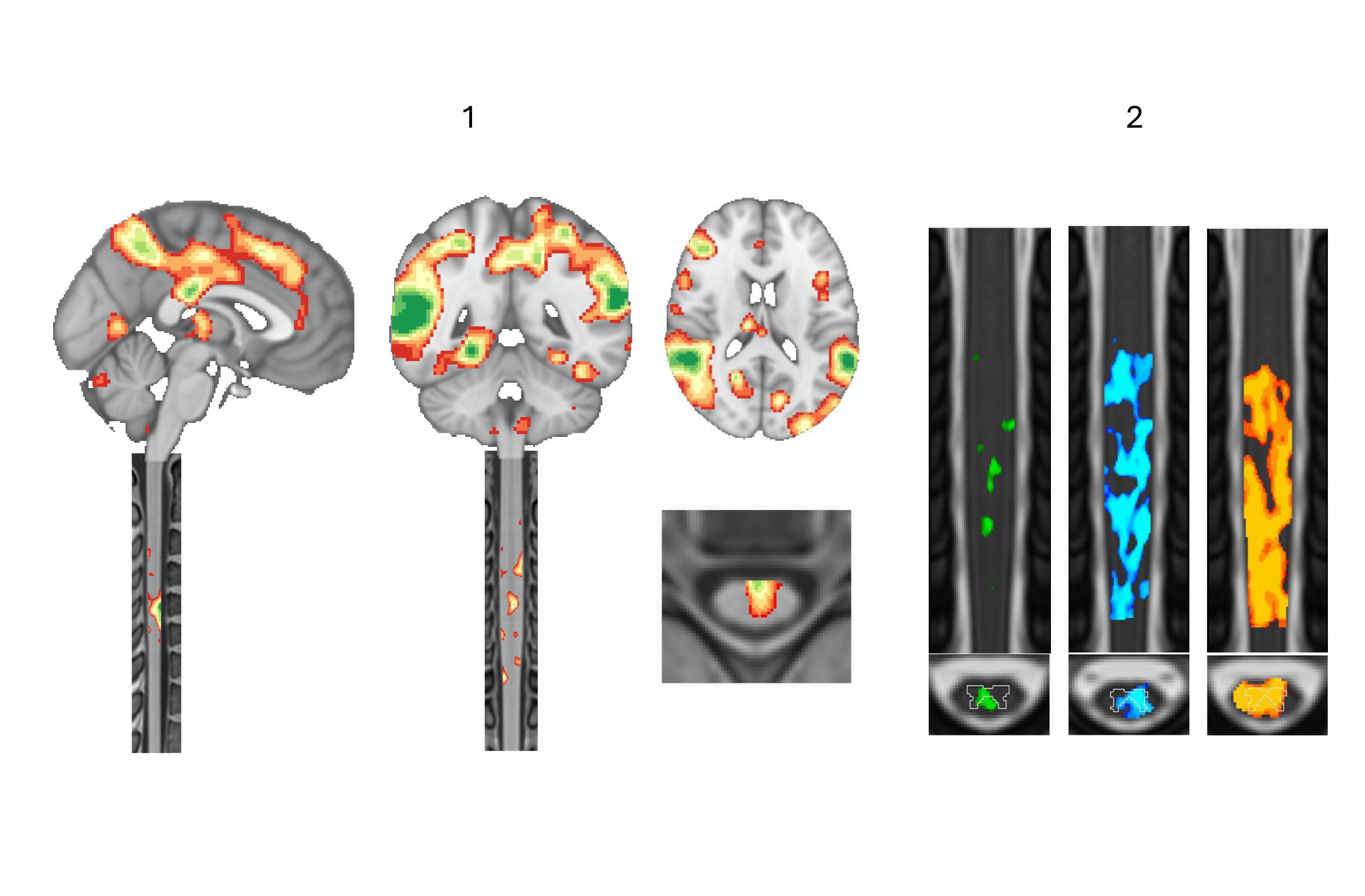
Functional MRI now maps activity beyond the brain. (1) Brain and cervical spinal cord imaged together in the same scan. (2) Spinal cord responses to magnetic pulses delivered to the motor cortex at increasing strength, from left to right: the stronger the pulse, the wider the activity spreads. EPFL/MIP:Lab – CC-BY-SA 4.0
<関連情報>
- https://actu.epfl.ch/news/using-brain-imaging-to-study-the-spinal-cord/?utm_source=chatgpt.com
- https://www.biorxiv.org/content/10.64898/2026.07.24.740278v1
- https://www.cell.com/neuron/fulltext/S0896-6273(20)30568-7
同時経頭蓋磁気刺激(TMS)と脊髄fMRIにより、脊髄回路の強度依存性変調が明らかになった Concurrent TMS and Spinal Cord fMRI Reveal Intensity-Dependent Modulation of Spinal Circuits
Ekansh Sareen, Rebecca Jones, Estelle Raffin, Friedhelm C. Hummel, Dimitri Van De Ville
bioRxiv Posted: July 28, 2026
DOI:https://doi.org/10.64898/2026.07.24.740278
Abstract
The spinal cord serves as a crucial relay for motor commands, yet the role of its local circuitry in sensorimotor integration remains poorly understood. Most non-invasive cortical stimulation studies, rely on electrophysiological readouts or inferred spinal function from corticospinal anatomy, leaving the downstream impact of cortical stimulation on spinal circuitry largely uncharted in vivo. Advances in spinal cord functional MRI (SC-fMRI) now enable spatially resolved imaging of segmental gray and white matter and their interactions with descending cortical inputs. Here, we introduce a multimodal framework that combines single-pulse transcranial magnetic stimulation (TMS) of the primary motor cortex with SC-fMRI to probe TMS-evoked spinal activity in humans. Using graded TMS intensities, we examined blood oxygenation level-dependent (BOLD) responses in the cervical spinal cord and asked how spinal activation depends on effective engagement of the descending motor system. Our findings reveal robust, intensity-dependent spinal BOLD responses aligned with descending pathways, with activation concentrated in expected territories such as the lateral corticospinal tract and ventral horn at segments innervating the stimulated hand muscle. By linking peripheral output to segment- and pathway-resolved spinal signals, these results demonstrate that concurrent TMS–SCfMRI can capture, in vivo, how cortical drive is expressed within human spinal circuitry and provide a new framework to measure spinal contributions to sensorimotor control with spatial specificity beyond traditional peripheral readouts.
安静時脊髄fMRIの動的機能的連結性により、微細な固有構造が明らかになる Dynamic Functional Connectivity of Resting-State Spinal Cord fMRI Reveals Fine-Grained Intrinsic Architecture
Nawal Kinany ∙ Elvira Pirondini ∙ Silvestro Micera ∙ Dimitri Van De Ville
Neuron Published: September 9, 2020
DOI:https://doi.org/10.1016/j.neuron.2020.07.024
Highlights
- The SpiCiCAP framework can delineate functional spinal circuits in fMRI data
- Components are revealed that are highly structured and in line with neuroanatomy
- Network organization emerges based on ascending and descending spinal neural pathways
- Pathway-specific patterns of temporal properties are highlighted
Summary
The neuroimaging community has shown tremendous interest in exploring the brain’s spontaneous activity using functional magnetic resonance imaging (fMRI). On the contrary, the spinal cord has been largely overlooked despite its pivotal role in processing sensorimotor signals. Only a handful of studies have probed the organization of spinal resting-state fluctuations, always using static measures of connectivity. Many innovative approaches have emerged for analyzing dynamics of brain fMRI, but they have not yet been applied to the spinal cord, although they could help disentangle its functional architecture. Here, we leverage a dynamic connectivity method based on the clustering of hemodynamic-informed transients to unravel the rich dynamic organization of spinal resting-state signals. We test this approach in 19 healthy subjects, uncovering fine-grained spinal components and highlighting their neuroanatomical and physiological nature. We provide a versatile tool, the spinal innovation-driven co-activation patterns (SpiCiCAP) framework, to characterize spinal circuits during rest and task, as well as their disruption in neurological disorders.