2026-11-11 北海道大学,大阪大学
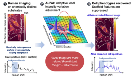
図1.Adaptive Local Intensity Variation Adjustment(ALIVA)補正によるハイドロゲル上培養細胞のラマンスペクトルの解析。①⾜場環境のラマン信号の混⼊、照明ムラを反映したラマン画像。②Toblerの地理学における第⼀原理「「近いものほど互いに関連が強い」」とベイズ最適化に基づく局所環境補正の概念図。③ALIVAによる局所環境補正後のラマン画像。
<関連情報>
- https://www.hokudai.ac.jp/news/2026/11/post-2499.html
- https://www.hokudai.ac.jp/news/pdf/261111_pr.pdf
- https://pubs.acs.org/ancham/article-abstract/doi/10.1021/acs.analchem.6c03142/5465914/Extracting-Single-Cell-Raman-Signature-in-Scaffold
ベイズ最適化を用いた局所背景補正による足場環境下のシングルセル固有のラマン特性を抽出する Extracting Single-Cell Raman Signature in Scaffold Environments through Bayesian-Optimized Local Background Correction
Jean-Emmanuel Clément; Zannatul Ferdous § ; Khalifa Mohammad Helal; Masumi Tsuda; Yasuaki Kumamoto; Menglu Li; Thomas Bocklitz; Jian Ping Gong; Katsumasa Fujita; Shinya Tanaka; Tamiki Komatsuzaki
Analytical Chemistry Published:November 06, 2026
DOI:https://doi.org/10.1021/acs.analchem.6c03142
Abstract
Despite the fact that a hydrogel provides a scaffold on which cells experience elevation of stemness marker expression in cell culture, technologies for real-time, noninvasive monitoring of biochemical states in these 3-dimensional (3D) systems remain limited. Recent advances in high-speed Raman imaging enable label-free biomolecular mapping with high spatial resolution and chemical specificity; however, spectral interference from scaffold polymers often obscures cellular features and complicates data interpretation. Here, we introduce a Bayesian-optimized background subtraction method that employs spatially adaptive Gaussian kernels to minimize scaffold contamination that may have spatial inhomogeneities. Applied to Raman images of murine C2C12 cells infused with the synovial sarcoma fusion gene SS18–SSX1 cultured on scaffold hydrogel and quartz substrates, our method improved the overlap of cellular spectral signatures across substrates and reduced unwanted variation within substrates, as confirmed both in the principal component space and in the wavenumber space. Compared to conventional preprocessing techniques that consider only the spectral dimension in background correction, our method is shown to be more effective in suppressing substrate features in cell spectra. As a result, the random forest classification of cells between the types of scaffold was primarily driven by cellular information, whereas conventional methods led to decisions dominated by substrate information. This approach is expected to advance Raman imaging for hydrogel-based cell culture analysis, enabling more accurate metabolic profiling of single cells and may, after further validation, be extended to more complex biological systems such as organoids in complex in situ-like environments.