2026-07-07 ピッツバーグ大学
<関連情報>
- https://news.engineering.pitt.edu/this-imaging-technique-shows-nerves-in-jaw-dropping-clarity/
- https://www.nature.com/articles/s44303-026-00167-6
ラット膝関節における神経支配を可視化するための高度な組織透明化および三次元イメージング手法 Advanced tissue clearing and three-dimensional imaging approaches to visualize neural innervation in the rat knee joints
Mairobys Socorro,Janak Gaire,Juliane Rolim de Lavor,Yenisel Cruz-Almeida,Robert M. Caudle,Reese Smith,Simon C. Watkins,Kyle D. Allen,RE-JOIN Consortium,Alan M. Watson & Alejandro J. Almarza
npj Imaging Published:13 May 2026
DOI:https://doi.org/10.1038/s44303-026-00167-6
Abstract
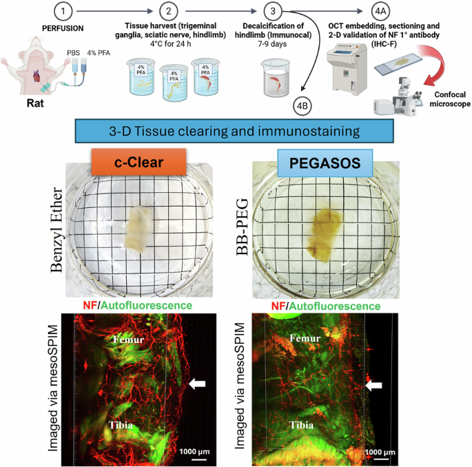
Tissue clearing techniques, combined with immunolabeling and three-dimensional (3-D) high-resolution imaging, have emerged as powerful tools for mapping the architecture of nerves that supply a specific tissue. However, despite significant advances in these techniques, visualizing nerve fibers within joint structures remains a technically challenging task. Moreover, most current protocols are optimized for use in mice, limiting their application in other animal species with greater tissue size, such as rats, which offer advantages for anatomical studies combined with behavioral models of sensory innervation and pain. Therefore, continued refinement of tissue clearing and imaging methods in rat tissue is essential for improving the resolution and translational relevance of joint innervation studies. In this work, we assessed and compared two tissue clearing protocols, a modified polyethylene glycol-associated solvent system (PEGASOS) and a newly developed hybrid method that combines CUBIC/3DISCO for tissue clearing (c-Clear), to visualize neurofilament-positive (NF+ ) nerve fibers in rat knees using 3-D fluorescence imaging. In summary, c-Clear resulted in a better option to detect NF+ fibers in the rat knee, whereas PEGASOS cleared tissues revealed greater autofluorescence within the muscle and bone marrow, compromising neurofilament visualization. Additionally, we highlight the importance of multi-angle imaging approaches when using cutting-edge light-sheet microscopy to capture the spatial context of neural innervation patterns within the complex knee microenvironment.