2026-09-28 東京科学大学
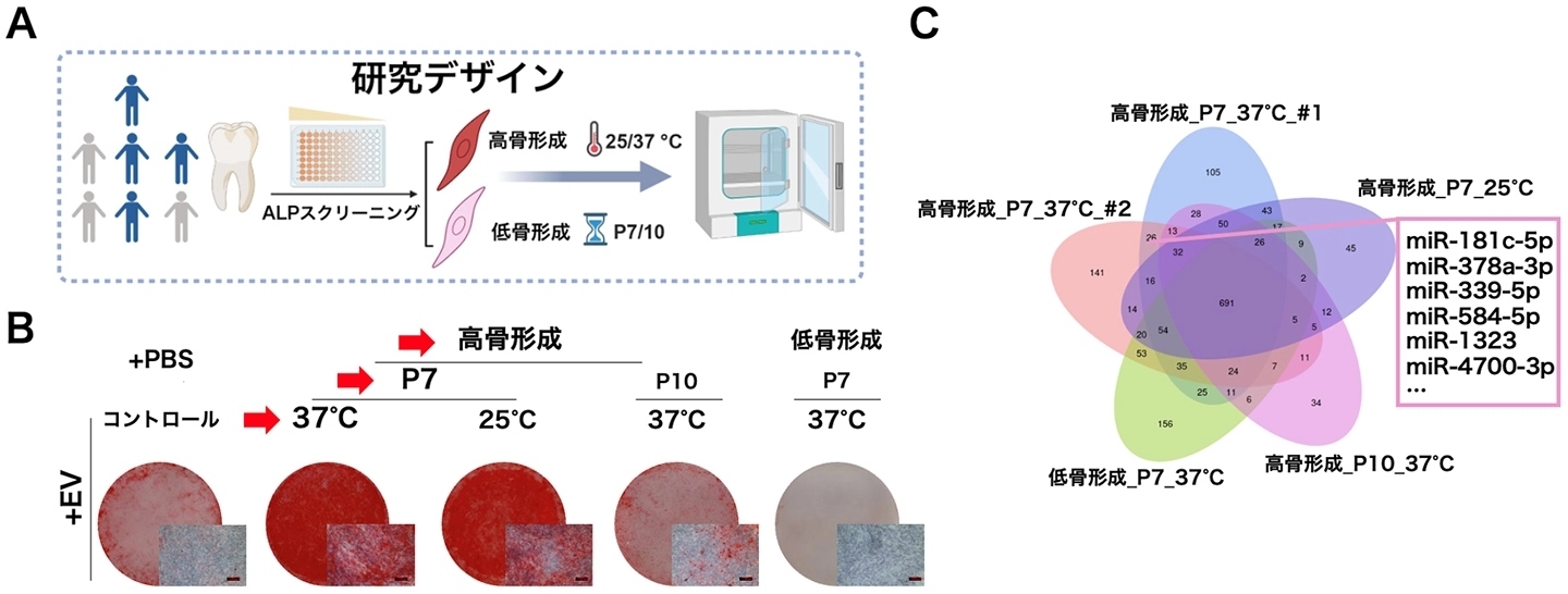
図1. 研究デザインとhPDL-MSC由来EVの性状・機能評価。
(A)研究デザインの模式図。(B)異なる継代数(P7、P10)および温度条件(37℃、25℃)で得たEVを処理したOsteo_High/Osteo_Low細胞由来EVを投与した際のアリザリンレッドS染色。
<関連情報>
- https://www.isct.ac.jp/ja/news/uy1u8k5wm5mu
- https://link.springer.com/article/10.1186/s12964-026-03153-y
- https://link.springer.com/article/10.1007/s12015-026-11160-2
細胞外小胞からの標的遺伝子miR-181cシグナル伝達は、RNF150-MAP3K5を介して歯周MSCの運命を制御する Targetome-defined miR-181c signaling from extracellular vesicles governs periodontal MSC fate via RNF150-MAP3K5
Jingyi Gao, Anhao Liu, Ye Yint Kaung Myint, Masahiro Hatasa, Supreda Suphanantachat Srithanyarat & Takanori Iwata
Cell Communication and Signaling Published:23 September 2026
DOI:https://doi.org/10.1186/s12964-026-03153-y
Abstract
Background
In regenerative settings, robust differentiation of human mesenchymal stromal cells (hMSCs) requires precise coupling between post-transcriptional regulation and kinase-driven pathways to achieve optimal therapeutic efficacy. miRNA-mediated regulation is widely implicated in stem cell fate control, yet its mechanistic contribution to lineage commitment remains incompletely defined.
Methods
Multi-conditioned periodontal ligament–derived hMSCs were developed as the regenerative model for alveolar bone. Extracellular vesicle (EV)-encapsulated miRNAs were delineated through microarrays. AGO2 RNA-immunoprecipitation sequencing with transcriptome profiling was integrated to establish the miRISC-associated targetome. Protein-protein interactions and signaling hierarchy were examined by Co-IP, WB, and IFC under knockdown/overexpression with pharmacological interventions. The translational efficacy of EV and miRNA was validated using a mouse ligature-induced periodontitis model, followed by µCT and histological analyses.
Results
EVs produced by hMSCs with higher intrinsic osteogenic capacity exhibited enhanced osteo-inductivity, traced to the enrichment of miR-181c. miR-181c was consistently upregulated during osteogenesis, with gain- and loss-of-function producing concordant effects. Mechanistically, the E3 ubiquitin ligase RNF150 emerged as the main target, whose repression reduced MAP3K5 ubiquitin-mediated proteolysis and enabled p38 activation. Local administration of EVs and miR-181c both promoted alveolar bone regeneration process in vivo.
Conclusion
Collectively, EV–miR-181c–RNF150–MAP3K5–p38 axis was proposed linking miRNA-mediated repression to kinase activation and lineage commitment. This work provides the framework for how EV-delivered miRNAs gate hMSCs’ fate decision, with significance for acellular therapeutic modalities in periodontal and skeletal tissues.
hsa-let-7c-3pはNT5Eを介してヒト歯周靭帯間葉系幹細胞/間質細胞の増殖から骨形成への移行を抑制する hsa-let-7c-3p Restrains the Proliferation-to-Osteogenesis Transition of Human Periodontal Ligament Mesenchymal Stem/Stromal Cells via NT5E
Ye Yint Kaung Myint, Anhao Liu, Jingyi Gao, Masahiro Hatasa, Supreda Suphanantachat Srithanyarat & Takanori Iwata
Stem Cell Reviews and Reports Published:02 June 2026
DOI:https://doi.org/10.1007/s12015-026-11160-2
Abstract
Small extracellular vesicles (sEVs) derived from mesenchymal stem/stromal cells (MSCs) contain microRNAs (miRNAs) that can modulate the cellular behavior of recipient cells. Human periodontal ligament mesenchymal stem/stromal cells (hPDL-MSCs) are a unique MSC population involved in periodontal tissue regeneration; however, the specific miRNAs that regulate their proliferative and differentiation–related properties remain incompletely understood. In this study, we profiled miRNAs enriched in sEVs secreted by highly proliferative human PDL-MSCs and identified hsa-let-7c-3p as a candidate regulator of hPDL-MSC behavior. Functional assays demonstrated that hsa-let-7c-3p enhanced proliferative and migratory behavior, reflected by an approximately 1.5-fold increase in metabolic activity and a 2-fold increase in migration, while suppressing osteogenic differentiation, with approximately 50% reductions in alkaline phosphatase activity and mineralization. To identify functionally relevant downstream targets, we intersected AGO2-RIP-seq–enriched transcripts, RNA-seq–defined genes altered by hsa-let-7c-3p overexpression, and in silico target predictions, identifying NT5E/CD73 as a candidate downstream target with experimental validation. Furthermore, NT5E knockdown partially phenocopied the effects of hsa-let-7c-3p overexpression on hPDL-MSC proliferation, migration, and osteogenic differentiation, supporting a functional link between hsa-let-7c-3p and NT5E/CD73. These findings highlight hsa-let-7c-3p as an underexplored sEV-associated 3p miRNA strand that restrains the proliferation-to-osteogenesis transition of hPDL-MSCs and identify NT5E/CD73 as a functionally relevant downstream target. Clinical trial number. Not applicable.