
2026-09-14 京都大学
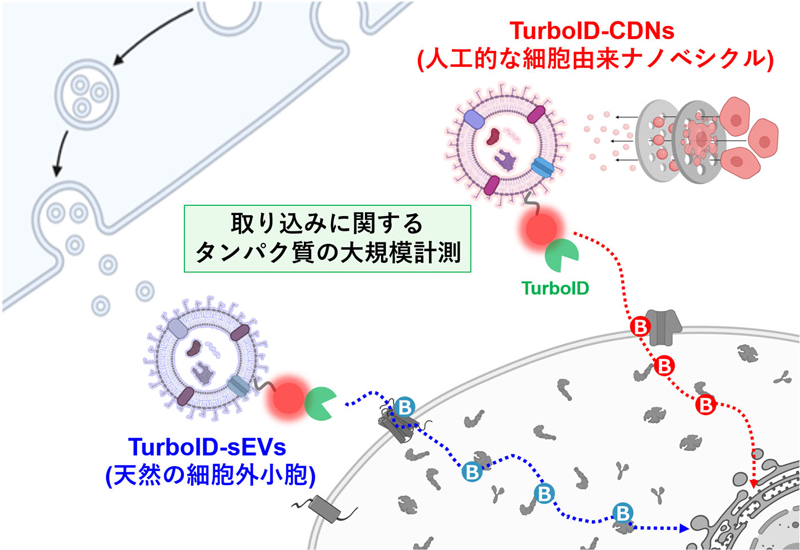
ナノ粒子の取り込み経路を調べる本研究の計測法。イラストはBioRender.com.で作成。
<関連情報>
- https://www.kyoto-u.ac.jp/ja/research-news/2026-09-14-1
- https://onlinelibrary.wiley.com/doi/10.1002/smtd.71014
近接プロテオミクスによる細胞由来ナノベシクルの細胞取り込み候補経路のマッピング Proximity Proteomics Maps Candidate Cellular Uptake Pathways for Cell-Derived Nanovesicles
Eisuke Kanao, Ryosuke Mizuta, Saki Tarao, Yukika Hara, Yuka Li, Kazunari Akiyoshi, Yasushi Ishihama, Koshi Imami, Yoshihiro Sasaki
Small Methods Published: 02 September 2026
DOI:https://doi.org/10.1002/smtd.71014
ABSTRACT
Although extracellular vesicles (EVs) facilitate selective molecular exchange between cells, their low yields and inherent heterogeneity limit translational applications. Cell-derived nanovesicles (CDNs), produced by mechanical extrusion of donor cells, offer a scalable alternative while retaining key membrane features of EVs. Yet how fabrication reshapes vesicle–cell communication remains poorly understood. Here, vesicle-side proteomics is integrated with TurboID proximity labeling of recipient-cell proteins to construct quantitative, dual-sided maps of CDN and small extracellular vesicle (sEV) interactions. Despite similar size and surface charge, CDNs displayed a substantially more diverse proteome and a broader repertoire of predicted uptake-associated signatures. Recipient-cell proximity proteomics further resolved distinct molecular cohorts. The CDN-associated cohort contained recipient-derived CALR and NCL, which are literature-linked to LRP1/CD91-associated efferocytic recognition and NCL-associated macropinocytic processes, respectively, whereas the sEV-associated cohort contained signatures consistent with HSPG-assisted docking and clathrin-mediated or CLIC/GEEC-related uptake. Together, these complementary datasets reveal molecular interfaces for extrusion-generated CDNs that are distinct from those of naturally secreted sEVs. Receptor dependence and the contributions of surface association and internalization require direct testing; however, the identified associations define specific mechanistic targets for further investigation. This dual-sided proteomic strategy establishes a quantitative framework for dissecting vesicle–cell communication and engineering membrane-based nanocarriers.

