2026-09-15 東京科学大学
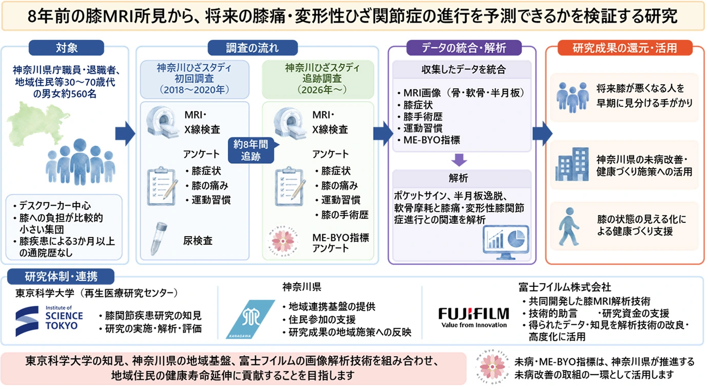
図1. 神奈川ひざスタディ追跡調査の概要
<関連情報>
MRI画像上の半月板脛骨靭帯ポケットは、内側膝関節変形性関節症の放射線学的重症度と関連している Meniscotibial ligament pockets on MRI are associated with radiographic severity of medial knee osteoarthritis
Hisako Katano,Nobutake Ozeki,Yusuke Nakagawa,Hideyuki Koga,Chiaki Okumura,Noriya Okanouchi,Jun Masumoto & Ichiro Sekiya
Scientific Reports Published:02 June 2026
DOI:https://doi.org/10.1038/s41598-026-52184-5
Abstract
Meniscotibial ligament dysfunction has been proposed as a factor contributing to medial meniscus extrusion (MME), a key feature of medial knee OA. This study investigated meniscotibial ligament pockets, defined as fluid-equivalent signals on MRI, as MRI findings that may reflect changes at the meniscotibial ligament insertion, including possible detachment. We examined their prevalence across Kellgren–Lawrence (KL) grades and their anatomical distribution. Participants from the Kanagawa Knee Study without lateral knee OA underwent spoiled gradient–recalled MRI. The “pocket sign,” defined as a fluid-equivalent signal between the medial tibial cortex and the ligament attachment on axial and coronal images, was used as an MRI finding potentially related to meniscotibial ligament detachment. Multivariable logistic regression evaluated associations between pocket presence and clinical factors. Pocket location was classified relative to the medial collateral ligament (MCL). A subset underwent surgical assessment during knee arthroplasty. Pocket prevalence increased with KL grade, from 40% in KL grade 0 to 100% in KL grade 4. Higher KL grade was independently associated with pocket presence (OR 2.56 per grade increase, p < 0.001). Age showed a limited and inconsistent association, whereas sex and BMI were not associated. In KL grade 0 knees, pockets were predominantly located anterior to the MCL and were associated with mild MME. Surgical findings supported the MRI results. Meniscotibial ligament pockets are present even in KL grade 0 knees and become more frequent with increasing radiographic severity. Their anterior predominance relative to the MCL suggests a characteristic distribution pattern. The pocket sign may serve as an MRI marker of meniscotibial ligament status.