2026-09-23 ペンシルベニア州立大学(Penn State)
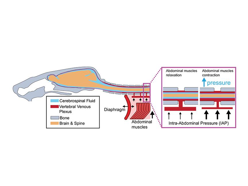
Schematic of mouse skull, brain, spinal column and abdominal muscles, showing how contraction of abdominal muscles could increase pressure in blood vessels connected to the brain through the spinal column. A new study by researchers at Penn State shows that the increased pressure creates ultrafast constrictions of major veins in the brain, momentarily increasing blood flow. Credit: Drew Lab / Penn State. Creative Commons
<関連情報>
- https://www.psu.edu/news/research/story/engaging-core-could-be-regulating-blood-flow-brain
- https://www.pnas.org/doi/10.1073/pnas.2604172123
- https://www.nature.com/articles/s41593-026-02279-z
腹圧によって引き起こされる脳内の超高速静脈および矢状静脈洞の収縮 Ultrafast venous and sagittal sinus constrictions in the brain driven by abdominal pressure
Qingguang Zhang, C. Spencer Garborg Noah Frank , +2 , and Patrick J. Drew
Proceedings of the National Academy of Sciences Published:September 23, 2026
DOI:https://doi.org/10.1073/pnas.2604172123
Abstract
Nearly all the blood supplying the cortex exits via the bridging veins (BVs) that drain into the superior sagittal sinus (SSS), making these vessels key chokepoints for cerebral blood flow. Using optical imaging in head-fixed mice, we found that the SSS, BVs, and some other pial veins exhibit ultrafast constrictions (<0.1 s) at the onset of locomotion, following whisker stimulation, and upon awakening from sleep. Constrictions of the BVs and the SSS were strongly correlated with abdominal muscle electromyography activity and were tightly correlated with respiration at rest. The rapid decrease in blood volume caused by venous constrictions resulted in spurious increases in fluorescence in mice expressing fluorescent reporter proteins, creating artifacts that could mimic functional signals. Venous constrictions with the same amplitude and dynamics could be generated in anesthetized mice by abdominal pressure application, showing that these constrictions were generated by mechanical coupling with the abdomen. Externally imposed abdominal pressures also drove a rapid but transient increase in blood flow. Unlike the pial and parenchymal microvasculature whose diameters are largely controlled by local signals, the diameters of SSS/BVs are dynamically controlled during behavior in part by abdominal muscle regulation of intracranial pressure, establishing a pathway for regulation of cerebral hemodynamics via mechanical coupling between the central nervous system and the viscera.
脳の動きは腹部との機械的な結合によって駆動される Brain motion is driven by mechanical coupling with the abdomen
C. Spencer Garborg, Beatrice Ghitti, Qingguang Zhang, Joseph M. Ricotta, Noah Frank, Sara J. Mueller, Denver I. Greenawalt, Kevin L. Turner, Ravi T. Kedarasetti, Marceline Mostafa, Hyunseok Lee, Francesco Costanzo & Patrick J. Drew
Nature Neuroscience Published:27 April 2026
DOI:https://doi.org/10.1038/s41593-026-02279-z
Abstract
The brain moves within the skull, but the drivers and consequences of this motion are not well understood. Here we visualized motion of the dorsal cortex relative to the skull in awake head-fixed mice using high-speed, multiplane two-photon microscopy. Brain motion was directed primarily rostrally and laterally, and was correlated tightly with locomotion, but not with respiration or the cardiac cycle. Specifically, brain motion was driven by abdominal muscle contractions that activate a hydraulic-like vascular connection between the nervous system and the abdominal cavity, and could similarly be induced by pressure applied to the abdomen. Model simulations suggest that brain motion may drive interstitial fluid through and out of the brain into the subarachnoid space, in the opposite direction of fluid flow seen during sleep. These results suggest that the brain is linked mechanically to the abdominal compartment, and that fluid flow in the brain could be coupled to body movements.