2026-10-08 オックスフォード大学
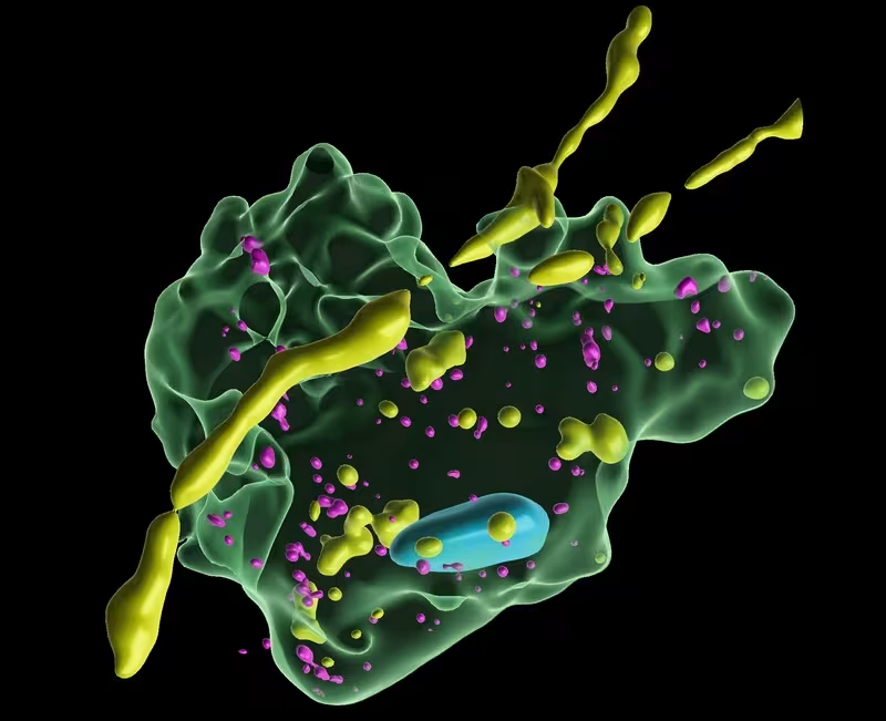
3D reconstruction of Airyscan super-resolution image of MG cocultured with DANs under fibril-seeded conditions, showing the presence of pSyn+ puncta and MAP2+ puncta in CD68+ MG. Image credit: Tofaris Group, University of Oxford.
<関連情報>
- https://www.ox.ac.uk/news/2026-10-06-brain-immune-cells-protect-neurons-in-parkinsons-disease-by-nibbling-harmful
- https://www.science.org/doi/10.1126/scitranslmed.adz9258
ヒトミクログリアはGPNMBを介したトロゴサイトーシスによって神経細胞内α-シヌクレイン凝集体を除去する Human microglia clear intraneuronal α-synuclein aggregates by GPNMB-mediated trogocytosis
Hung-Ju Chueh, Antigoni Katsikoudi, Ana Aragón-González, Chor Lai Lam, […] , and George K. Tofaris
Science Translational Medicine Published:7 Oct 2026
DOI:https://doi.org/10.1126/scitranslmed.adz9258
Abstract
Microglia are the primary immune cells of the brain, but their role in Parkinson’s disease is not fully understood. Chronic microglial activation is toxic to neurons, but in the early stages of pathology, microglia also exert beneficial functions. We used induced pluripotent stem cell (iPSC)–derived coculture models to investigate how human microglia respond to α-synuclein aggregates that form de novo inside human dopaminergic neurons with α-synuclein gene triplication or are triggered by fibrils. We found that microglia cleared Ser129-phosphorylated α-synuclein aggregates through a contact-dependent mechanism. This process was fine-tuned by sensing (P2RY12) and inhibitory (CD22) signals and involved selective phagocytosis of neuronal subcompartments (trogocytosis), independently of TREM2 or phosphatidylserine. In the presence of intraneuronal α-synuclein aggregates, microglia exhibited morphological and transcriptional changes indicative of activation. Using single-cell sequencing, we identified a cluster of disease-associated microglia (DAM) responsible for this beneficial phenotype and the Parkinson’s disease GWAS candidate glycoprotein nonmetastatic melanoma protein B (GPNMB) as a key effector of aggregate clearance by microglial lysosomes. GPNMB interacted with Ser129-phosphorylated α-synuclein in iPSC-derived microglia exposed to aggregate-laden dopaminergic neurons and was up-regulated in substantia nigra microglia of individuals with incidental Lewy bodies or Parkinson’s disease. Microglia-specific GPNMB knockdown using CRISPRi reduced intraneuronal aggregate clearance. GPNMB-mediated trogocytosis was negatively regulated by an autocrine IL-10 signaling loop, whereas IL-10 receptor blockade enhanced the phagocytic response. Together, our study identifies a subtype of human microglia capable of removing intraneuronal aggregates and potential therapeutic targets.