2026-11-16 カリフォルニア大学ロサンゼルス校(UCLA)
カリフォルニア大学ロサンゼルス校(UCLA)を中心とする国際研究チームは、胎児期から出生後の乳児期にかけて、ヒトの大脳基底核とその周辺に存在する神経細胞がどのように発達するかを詳細に示す分子アトラスを構築した。研究では、妊娠中期から出生後7か月までの発達段階にあるヒト脳9例から、単一細胞核メチル化・3次元ゲノム解析法(snm3C-seq)により15万5,000個以上の細胞核を解析した。既存データと統合した解析対象は約28万7,000細胞に達した。
その結果、神経細胞の種類によって成熟する時期が異なることが明らかになった。特に、大脳基底核の主要な神経細胞である有棘投射ニューロンは、特徴的な同期したエピゲノム変化を経て成熟することが示された。また、オリゴデンドロサイトの分化過程では、出生後に一時的な3次元クロマチン構造の変化が生じることも確認された。さらに、精神疾患の遺伝学的データとの比較から、有棘投射ニューロンには統合失調症と関連する遺伝的リスクが強く集積し、一部の細胞亜型では双極性障害との関連も示された。本アトラスは、脳の正常な発達過程を基準として、神経・精神疾患に関係する細胞種や発症に関わる発達時期を研究するための基盤となる。
Mohammad S. Baig, Luo Lab
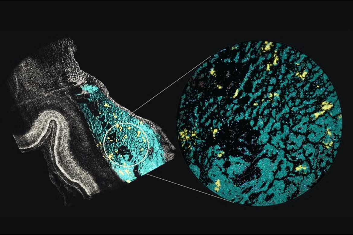
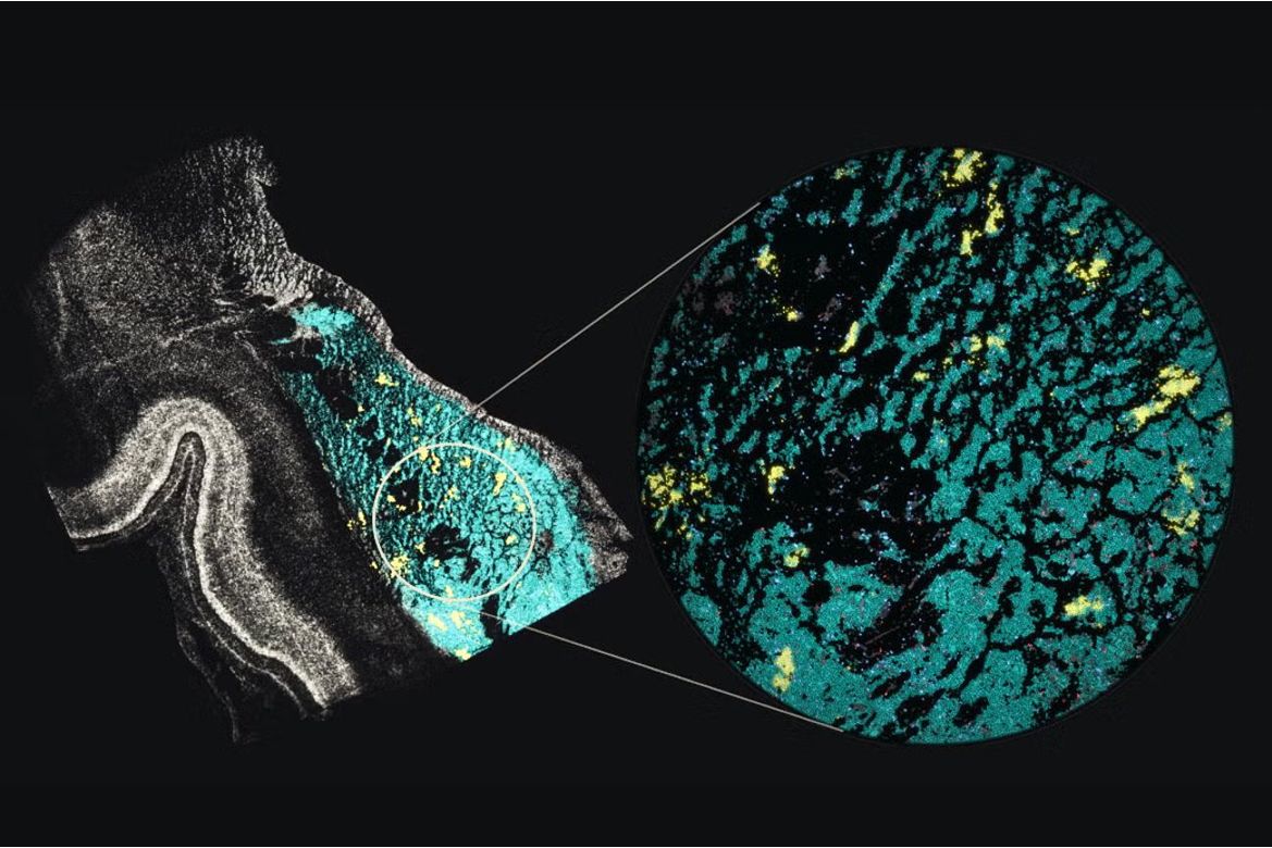
A view of the developing human basal ganglia shows small islands of striosome cells (yellow) surrounded by matrix cells (blue). Both are subtypes of spiny projection neurons, whose development and gene regulation are mapped in the new atlas.
<関連情報>
- https://newsroom.ucla.edu/stories/womb-world-new-atlas-traces-development-key-human-brain-cells
- https://www.cell.com/cell/fulltext/S0092-8674(26)01219-5
発達中のヒト大脳基底核に関する単一細胞・空間情報を統合した3次元マルチオミクスアトラス
(A single-cell and spatial 3D multi-omic atlas of developing human basal ganglia)
Matthew G. Heffel, Heng Xu, Oier Pastor-Alonso, Xinzhe Li, Mohammad S. Baig, Rayyan Irfan Ghoor, Runjia Li, Colin Kern, Joonho Kum, Yi Zhang, Jon Paino, Gaurav Singh Rathore, Min Jen Tsai, Chu-Yi Tai, Grant Tucker, Zoey Zhao, Angie Hou, Zachary von Behren, Mohini Bhade, Siqian Li, Kadellyn Sandoval, Jessica Scholes, Felicia Codrea, Jeffrey Calimlim, Emma K. Liao, Gwyneth Leung, JaeYeon Kim, Alexandra LeFevre, Robert Johnson, Thomas Blanchard, Eleazar Eskin, Jonathan Flint, Jennifer A. Cotter, Bogdan Pasaniuc, Bogdan Bintu, Quan Zhu, Eran A. Mukamel, Jason Ernst, Mercedes F. Paredes, Chongyuan Luo
Cell, Volume 189, Issue 23, pp. 7550–7570.e11, 2026 Published: 12 November 2026
DOI:https://doi.org/10.1016/j.cell.2026.10.008

Graphical abstract (CC BY 4.0)
This study constructed a single-cell and spatial 3D multi-omic atlas to characterize the specification and maturation of the developing human basal ganglia. The researchers integrated single-nucleus methylation and chromosome conformation capture sequencing (snm3C-seq), spatial transcriptomics, and single-molecule chromatin and RNA imaging. The analysis revealed distinct developmental timelines among subregions of the ganglionic eminences (GEs), transient structures that give rise to basal ganglia neurons and inhibitory interneurons. Spiny projection neurons (SPNs), the principal neuronal population of the basal ganglia, acquired distinct subtypes through synchronized three-dimensional epigenomic remodeling, whereas other neuronal populations exhibited asynchronous maturation. The study also identified a transient short-range chromatin state during oligodendrocyte differentiation in infancy, demonstrating that chromatin conformations associated with terminal cell fates can also occur temporarily. In addition, the temporal cortex showed faster epigenomic maturation than other cortical regions during the examined developmental period. Finally, SPNs were found to be enriched for genetic risk associated with neuropsychiatric disorders. These findings provide an integrated framework for understanding cell-type-specific developmental trajectories and dynamic three-dimensional epigenomic changes in the human brain.