2026-09-04 東京科学大学
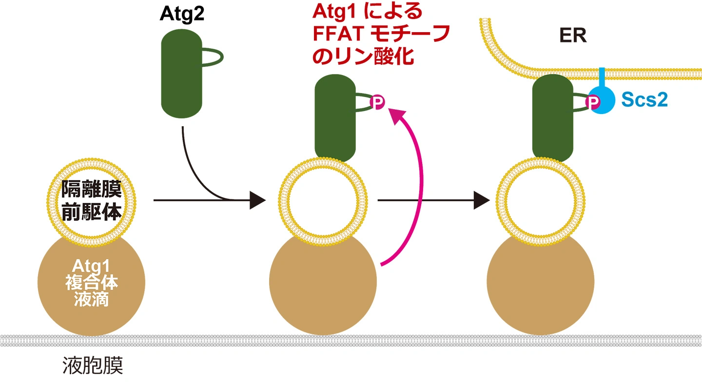
図. Atg1によるリン酸化を介したAtg2の小胞体認識機構
<関連情報>
オートファゴソーム形成中のAtg2とERの結合の時空間制御 Spatiotemporal control of Atg2 association with the ER during autophagosome formation
Tetsuya Kotani, Haruki Tanabe, Shinri Kitta, +8 , and Hitoshi Nakatogawa
Proceedings of the National Academy of Sciences Published:July 29, 2026
DOI:https://doi.org/10.1073/pnas.2606606123
Abstract
Autophagy degrades various intracellular components by sequestering them within membrane vesicles called autophagosomes and delivering them to lysosomes or vacuoles. Previous studies have revealed that the conserved, bridge-like lipid transfer protein Atg2 tethers autophagosome precursors to the endoplasmic reticulum (ER) and mediates lipid supply from the ER to drive their expansion into autophagosomes. However, how Atg2 docks onto the ER has remained unclear. Here, we show in Saccharomyces cerevisiae that Atg2 interacts with the ER-resident VAP family protein Scs2. This interaction is mediated by a phospho-FFAT motif in Atg2 and the major sperm protein domain of Scs2 and enhanced by phosphorylation of the motif by the autophagy-initiating kinase Atg1, which is activated at the autophagosome formation site upon autophagy induction. This interaction cooperates with the N-terminal region of Atg2, which contains a weakly amphipathic helix, to mediate Atg2 association with the ER. Thus, the Atg2–Scs2 interaction functions as a spatiotemporal switch that controls Atg2–ER association. We also show that mammalian ATG2 interacts with the VAP-like proteins MOSPD1 and MOSPD3 to promote autophagosome formation. Collectively, this study reveals a conserved mechanism that initiates lipid transfer during autophagosome formation.