2026-07-07 バッファロー大学(UB)
<関連情報>
- https://www.buffalo.edu/news/releases/2026/07/MS-scans-grey-matter-AI.html
- https://www.nature.com/articles/s43856-026-01683-7
多重コントラスト後処理と深層学習を用いた多発性硬化症MRIデータセットにおける皮質病変の定量化 Quantifying cortical lesions in multiple sclerosis MRI datasets using multi-contrast post-processing and deep learning
Michael G. Dwyer,Niels Bergsland,Alexander Bartnik,Dejan Jakimovski,Samantha Noteboom,Menno M. Schoonheim,Martijn D. Steenwijk,Jinglan Pei,David Clayton & Robert Zivadinov
Communications Medicine Published:07 July 2026
DOI:https://doi.org/10.1038/s43856-026-01683-7
Abstract
Background
Multiple sclerosis (MS) is a chronic neurological disease affecting both white and gray matter of the central nervous system. Despite the well-established involvement of cortical lesions in MS, feasibility limitations in their visualization on typical magnetic resonance imaging (MRI) protocols prevent their evaluation in nearly all clinical trials. Recently, several post-processing methods, including synthetic contrasts and artificial intelligence (AI)-based approaches, have shown potential for enhancing cortical lesion detection on conventional MRI data. These methods have the potential to reanalyze existing clinical-trial data to answer key mechanistic questions about both MS development and about treatment effects.
Methods
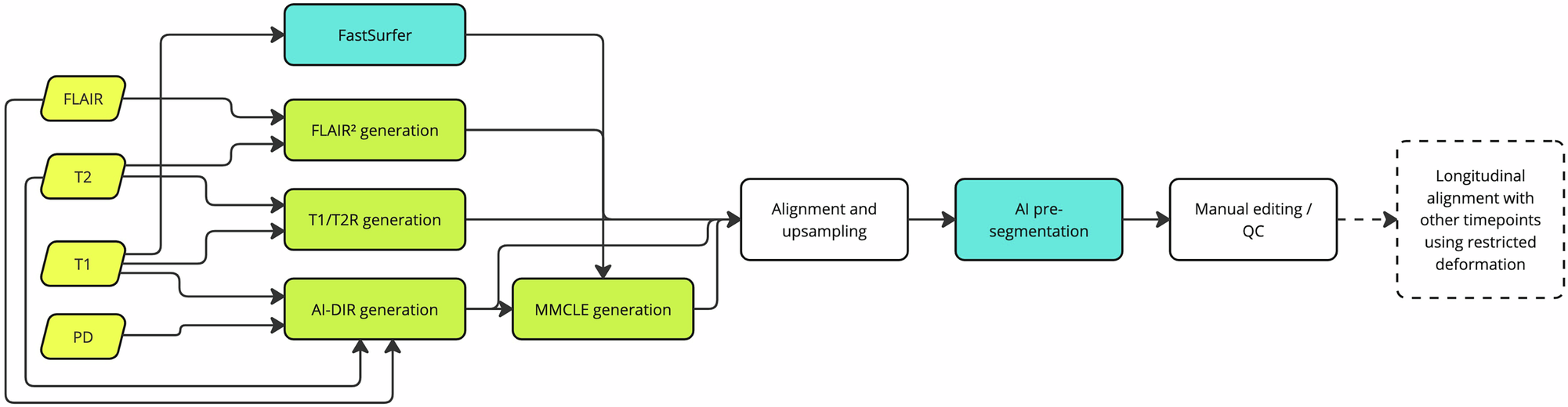
We sought to evaluate the feasibility of combining and extending existing methods into a unified framework for analysis using the data from the large, multicenter, phase 3 ORATORIO trial (full n = 732, age=44.6 ± 8.0; development subset n = 80, age=46.6 ± 7.1). We specifically evaluated three of the most promising of them – fluid-attenuated inversion recovery squared (FLAIR2), T1/T2 ratio, and artificial intelligence-derived double inversion recovery (AI-DIR) – and introduced a new combined contrast called multi-modal cortical lesion enhanced (MMCLE). We also harnessed transformer-based semantic segmentation to improve automated detection and delineation of these lesions.
Results
At baseline, we detected 14.8 + /−20.72 lesions per participant, with 86.0% true positive rate and 8.4% false positive rate across subjects for blinded MMCLE, using simultaneous review of all contrasts as the reference. High reproducibility was observed across field strengths and acquisition types (ICC 88.8-92.5%).
Conclusions
We confirmed that cortical lesions can be clearly visualized and quantified with these methods. Using deep learning, we also confirmed that the simultaneous use of multiple contrasts improves quantification.