2026-09-30 京都産業大学
<関連情報>
- https://www.kyoto-su.ac.jp/news/news-003541.html
- https://www.kyoto-su.ac.jp/mt_uploads/20260930_press02.pdf
- https://www.pnas.org/doi/10.1073/pnas.2627537123
クライオ電子顕微鏡解析によって明らかになった二成分毒素ポアの段階的成熟機構Step-by-step maturation mechanism of the binary toxin pore revealed by cryo-EM analysis
Tomohito Yamada, Ren Nakanishi, Yukihiko Sugita, +3 , and Hideaki Tsuge
Proceedings of the National academy of Sciences Published:September 28, 2026
DOI:https://doi.org/10.1073/pnas.2627537123
Abstract
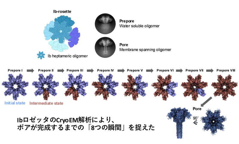
Membrane pore-forming proteins (PFPs) form ring-shaped membrane-translocating oligomers on membranes, contributing to infection, immunity, and cell death functions. Binary toxins produced by some bacteria consist of an enzymatic component that acts as a toxin and a membrane-binding component that forms a pore that delivers the enzymatic component into target cells. Cryoelectron microscopy (cryo-EM) has advanced our understanding of these translocation mechanisms by revealing several binary toxin complexes’ structures. However, the mechanisms underlying the initial pore formation remain unclear. We determined the structures of several oligomeric forms of the membrane-binding component Ib of the iota toxin from Clostridium perfringens at various stages of pore formation. Structural comparisons revealed how the symmetrically arranged soluble oligomer (prepore) asymmetrically matures into a transmembrane oligomer (pore). These findings enhance our understanding of mechanisms of PFP and provide a structural basis for developing nanodevices using membrane pores.