2023-03-16 ノースカロライナ州立大学(NCState)
米国ノースカロライナ州立大学の生物科学教授であるアン・ロス氏は、新たな法科学の研究が乳幼児の骨がどのように腐敗するかに光を当て、若い人の遺骸が特定の場所にどれくらい長くあったか、またDNAや他の組織検体を収集するために最適な骨がどれかを決定するのに役立つと述べています。
この研究では、人間の遺体に対する類似物として広く使用される家畜の豚の遺体を使用し、2年間にわたって乳幼児の遺体がどのように腐敗するかを調査しました。調査結果は、医療・法的コミュニティに若い人々への正義と、少しの閉鎖感をもたらすことが期待されます。
<関連情報>
温帯環境における幼若骨と胎児骨の異化のタイミングと範囲に関する研究 Investigating the Timing and Extent of Juvenile and Fetal Bone Diagenesis in a Temperate Environment
Amanda R. Hale and Ann H. Ross
Biology Published: 3 March 2023
DOI:https://doi.org/10.3390/biology12030403

Simple Summary
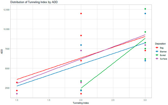
Understanding bone diagenesis, or alteration, in juvenile and fetal remains has important implications for forensic science. First, it can suggest information about the deposition of the remains and a possible postmortem interval (PMI). Second, it can assist in evaluating bone integrity and the potential for molecular testing of these remains for forensic purposes. This study investigates how early bone diagenesis is observed in fetal and juvenile mammalian remains as well as differences in degradation based on the deposition of the remains (e.g., blanket wrapping, shallow burial, etc.). We found that there were differences in the extent of bone diagenesis between depositions, with bagged remains exhibiting relatively less degradation over time than the other three depositions, while buried remains exhibited the greatest extent of degradation over time. However, all the remains showed bone diagenesis regardless of time of interment or deposition, with all remains exhibiting alteration as early as three months. This is consistent with adult remains, although the presentation of alteration differs and is likely related to developmental differences between subadult and adult bone.
Abstract
It is well understood that intrinsic factors of bone contribute to bone diagenesis, including bone porosity, crystallinity, and the ratio of organic to mineral components. However, histological analyses have largely been limited to adult bones, although with some exceptions. Considering that many of these properties are different between juvenile and adult bone, the purpose of this study is to investigate if these differences may result in increased degradation observed histologically in fetal and juvenile bone. Thirty-two fetal (n = 16) and juvenile (n = 16) Sus scrofa domesticus femora subject to different depositions over a period of two years were sectioned for histological observation. Degradation was scored using an adapted tunneling index. Results showed degradation related to microbial activity in both fetal and juvenile remains across depositions as early as three months. Buried juvenile remains consistently showed the greatest degradation over time, while the blanket fetal remains showed more minimal degradation. This is likely related to the buried remains’ greater contact with surrounding soil and groundwater during deposition. Further, most of the degradation was seen in the subendosteal region, followed by the subperiosteal region, which may suggest the initial microbial attack is from endogenous sources.


