2023-08-17 ヒューストン大学(UH)

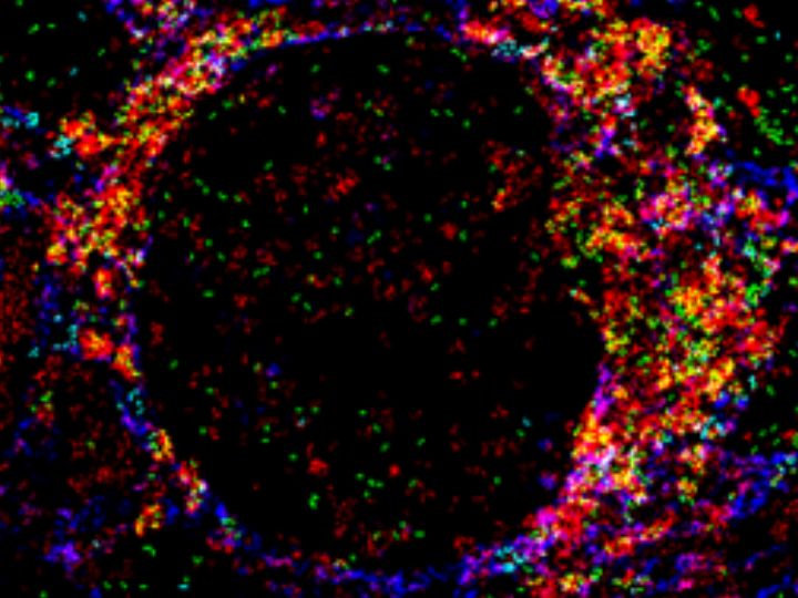
Photo of kidney glomeruli, color coded for different types of cells, taken by imaging mass cytometry, which can showcase the presence of as many as 37 different proteins simultaneously, a significant leap beyond the traditional approach, which allowed the examination of only 1-3 distinct proteins within a specific tissue.
◆LNは腎臓の重篤な炎症であり、ループス患者の死因の主要な要因です。IMCは、人間の組織内に37種類の異なるタンパク質の存在を同時に示すことができ、従来の方法では特定の組織内で1〜3つの異なるタンパク質のみを調査できる制限を大幅に超えるものです。IMCは、機械学習アルゴリズムと組み合わせて、ヒトの腎臓の細胞構造を特徴づけ、細胞タイプを区別し、疾患の新しいマーカーを同定するために使用されています。
<関連情報>
- https://uh.edu/news-events/stories/2023/august-2023/08172023-mohan-titus-mass-cytometry.php
- https://www.sciencedirect.com/science/article/abs/pii/S152166162300476X
50プレックスイメージングマスサイトメトリープロテオミクスで明らかになった増殖性ループス腎炎の分子構造 Molecular architecture of proliferative lupus nephritis as elucidated using 50-plex imaging mass cytometry proteomics
Anto Sam Crosslee Louis Sam Titus, Ying Tan, Phuongthy Tran, Julius Lindblom, Maryann Ivbievbiokun, Yitian Xu, Junjun Zheng, Ioannis Parodis, Qi Cai, Anthony Chang, Shu-Hsia Chen, Minghui Zhao, Chandra Mohan
Clinical Immunology Available online: 27 July 2023
DOI:https://doi.org/10.1016/j.clim.2023.109713
Abstract
Due to unique advantages that allow high-dimensional tissue profiling, we postulated imaging mass cytometry (IMC) may shed novel insights on the molecular makeup of proliferative lupus nephritis (LN). This study interrogates the spatial expression profiles of 50 target proteins in LN and control kidneys. Proliferative LN glomeruli are marked by podocyte loss with immune infiltration dominated by CD45RO+, HLA-DR+ memory CD4 and CD8 T-cells, and CD163+ macrophages, with similar changes in tubulointerstitial regions. Macrophages are the predominant HLA-DR expressing antigen presenting cells with little expression elsewhere, while macrophages and T-cells predominate cellular crescents. End-stage sclerotic glomeruli are encircled by an acellular fibro-epithelial Bowman’s space surrounded by immune infiltrates, all enmeshed in fibronectin. Proliferative LN also shows signs indicative of epithelial to mesenchymal plasticity of tubular cells and parietal epithelial cells. IMC enabled proteomics is a powerful tool to delineate the spatial architecture of LN at the protein level.

