2026-07-24 東京大学
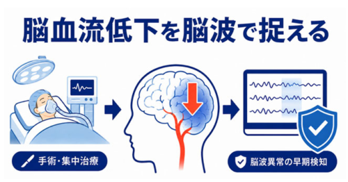
本研究結果が示す脳波モニタリングの可能性
<関連情報>
- https://www.h.u-tokyo.ac.jp/press/20260724-2.html
- https://www.h.u-tokyo.ac.jp/press/__icsFiles/afieldfile/2026/07/24/release_20260724-2.pdf
- https://www.bjanaesthesia.org/article/S0007-0912(26)00411-3/abstract
時間周波数表現は、急性脳低灌流時の見かけ上の脳波徐波化を形成する
Time–frequency representation shapes apparent electroencephalographic slowing during acute cerebral hypoperfusion
Ryo Wakabayashi Send email to wakabayashir@g.ecc.u-tokyo.ac.jp ∙ Yuka Tatematsu ∙ Seiichi Azuma ∙ Masaaki Asamoto ∙ Kanji Uchida
British Journal of Anaesthesia Published: July 23, 2026
DOI:https://doi.org/10.1016/j.bja.2026.05.017
During cerebral hypoperfusion, the processed EEG typically progresses from spectral slowing to suppression as the severity of hypoperfusion increases.1,2 Nonetheless, the ability of processed EEG indices to detect acute cerebral hypoperfusion remains unclear.3 Time–frequency displays are increasingly used in perioperative EEG monitoring and can depict evolving EEG slowing during acute cerebral hypoperfusion.4,5 However, these displays are analytic constructions shaped by proprietary method-specific assumptions.6 For example, the multitaper method (MTM) generates stable spectral estimates through fixed-window averaging with orthogonal tapers but can temporally smooth abrupt transitions over short timescales.7 In contrast, the Hilbert–Huang transform (HHT) provides adaptive instantaneous frequency–energy representations derived from intrinsic mode functions and can capture short-lived non-stationary changes more sharply.8 We therefore hypothesised that EEG slowing during acute cerebral hypoperfusion would be represented differently by these contrasting analytic frameworks.