2026-08-04 東京科学大学
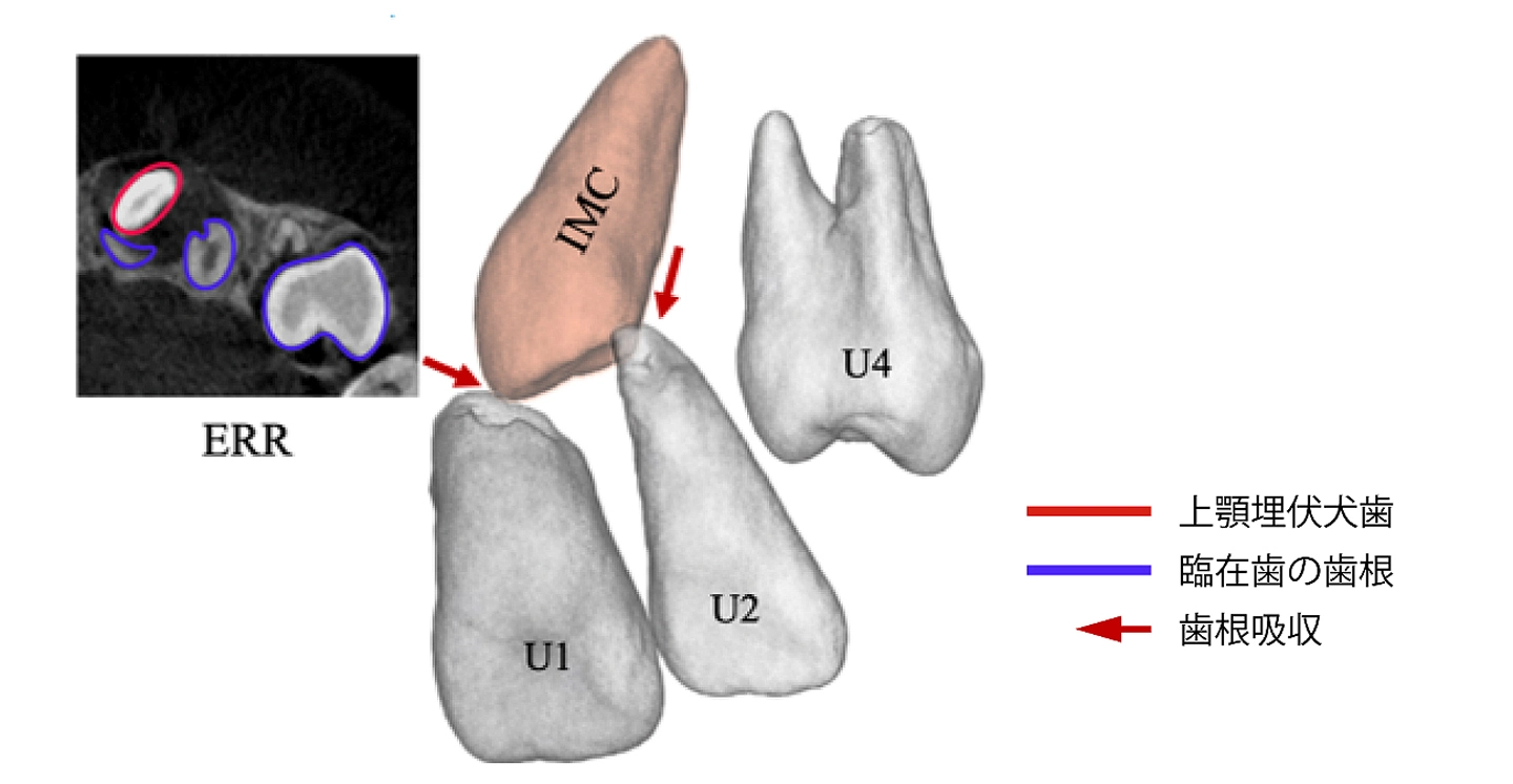
図1. 上顎埋伏犬歯による隣在歯の歯根吸収の一例。
上顎埋伏犬歯が隣在歯に接近することで、歯根吸収が生じています。CBCT画像では、その様子を明瞭に確認できます。
【略語】IMC:上顎埋伏犬歯、U1:上顎中切歯、U2:上顎側切歯、U4:上顎第一小臼歯
<関連情報>
- https://www.isct.ac.jp/ja/news/80kccitlhf7q
- https://link.springer.com/article/10.1186/s40510-026-00638-x
隣接歯の歯根形成の進行と埋伏上顎犬歯に伴う外部歯根吸収との関係:後向き横断的CBCT研究 Relationship between root formation progress of adjacent teeth and external root resorption associated with impacted maxillary canines: a retrospective cross-sectional CBCT study
Yukari Mizoguchi,Yuji Ishida,Aiko Ishizaki-Terauchi,Chiyo Shimizu-Tomoda & Takashi Ono
Progress in Orthodontics Published:14 July 2026
DOI:https://doi.org/10.1186/s40510-026-00638-x
Abstract
Background
This study aimed to clarify the involvement of the root formation stage and apical foramen diameter (AFD) of adjacent teeth in the occurrence of external root resorption (ERR) associated with impacted maxillary canines (IMCs), and to identify novel risk indicators for more accurate clinical prediction.
Methods
Forty patients with IMCs (58 canines) who underwent cone-beam computed tomography (CBCT) between May 2017 and October 2022 were retrospectively analyzed. The developmental status was assessed using the root formation stage and AFD of the central incisor (U1), lateral incisor (U2), and first premolar (U4). Additionally, spatial factors, including inter-root distance and the 3D position of the IMC, were evaluated in relation to ERR.
Results
ERR was detected in 72.5% of patients, affecting 37.1% of all examined adjacent teeth. Among the affected teeth, U2 showed the highest incidence (65.3%), followed by U1 (28.6%) and U4 (6.1%). In exploratory tooth-level comparisons, AFD was smaller in the ERR group for U1 and U4. In the supplementary affected-side-level mixed-effects analysis, no statistically significant independent association was demonstrated between maximum AFD and side-level ERR occurrence.
Conclusions
Developmental indicators, including AFD, may provide clinically relevant information when assessing ERR associated with IMCs. These indicators should be interpreted as potential modifying factors in conjunction with established spatial relationships rather than as independent predictive markers.