2026-11-02 東京大学,理化学研究所
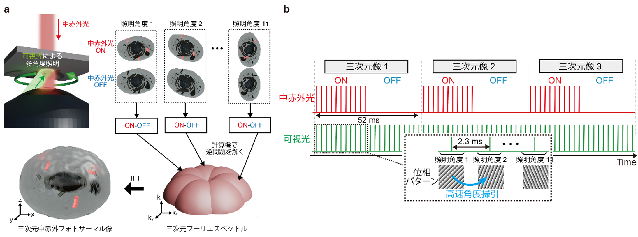
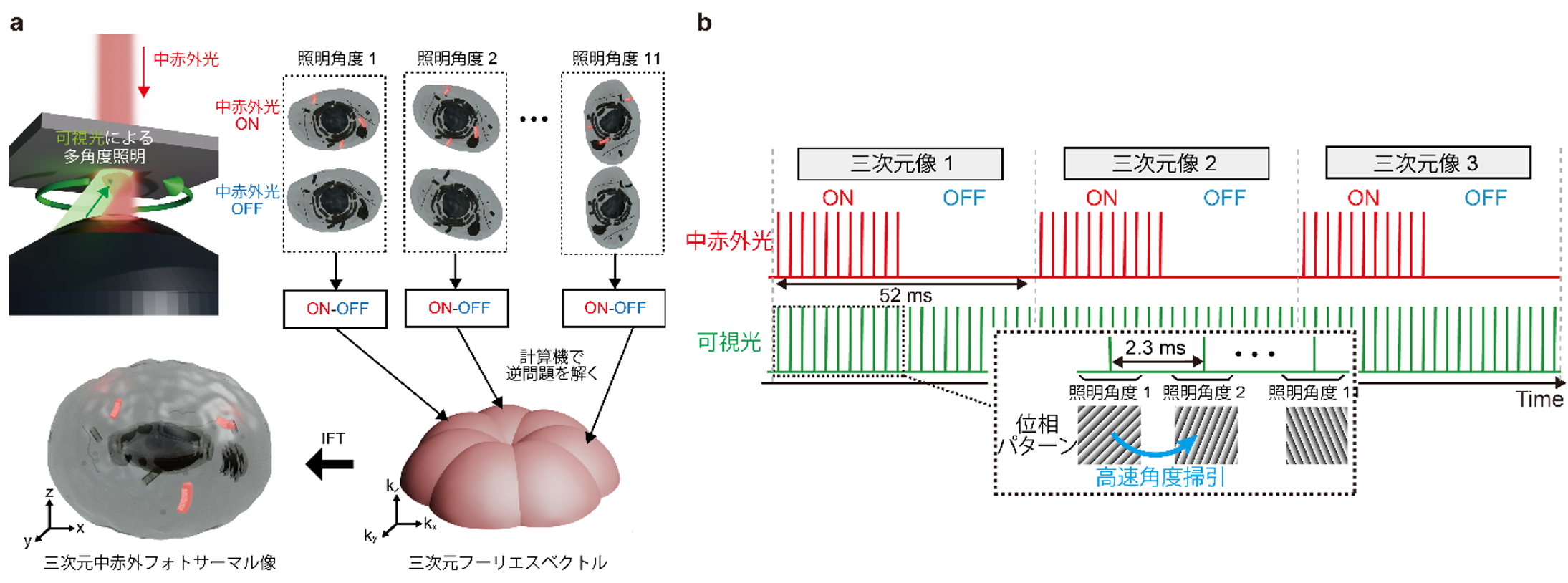
図1:ビデオレート三次元中赤外フォトサーマル顕微鏡の原理
<関連情報>
- https://www.k.u-tokyo.ac.jp/information/category/press/0030898.html
- https://www.nature.com/articles/s41467-026-78699-z
中赤外線光熱光学回折トモグラフィーによるビデオレート体積化学イメージング Video-rate volumetric chemical imaging via mid-infrared photothermal optical diffraction tomography
Masato Fukushima, Keiichiro Toda, Yusei Sugawara, Shotaro Kawano & Takuro Ideguchi
Nature Communications Published:30 October 2026
DOI:https://doi.org/10.1038/s41467-026-78699-z
Abstract
Label-free vibrational microscopy provides chemically specific access to cellular structures, yet quantitative volumetric imaging of intracellular dynamics remains challenging on subsecond timescales. Here, we demonstrate video-rate mid-infrared photothermal optical diffraction tomography (MIP-ODT) by combining high photothermal sensitivity with rapid angular scanning. The system achieves 19.2 volumes per second, nearly 400 times faster than previous MIP tomography implementations, with a signal-to-noise ratio exceeding 70 and no temporal averaging. We track lipid droplets in living cells and quantify anomalous diffusion from three-dimensional trajectories, revealing heterogeneous intracellular transport behaviors obscured in two-dimensional measurements. Rapid mid-infrared wavenumber sweeping further enables broadband multi-wavenumber volumetric chemical imaging across a 300 cm⁻¹ spectral window within 1 s. These results establish MIP-ODT as a platform for high-speed, label-free volumetric chemical imaging of single cells and quantitative investigation of dynamic intracellular organization.